. 2015 May 26;112(21):6688-93.
doi: 10.1073/pnas.1421699112. Epub 2015 May 11.

Human caspase-4 mediates noncanonical inflammasome activation against gram-negative bacterial pathogens

Affiliations

Human caspase-4 mediates noncanonical inflammasome activation against gram-negative bacterial pathogens

Cierra N Casson et al. Proc Natl Acad Sci U S A. .

Abstract

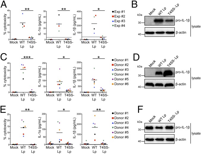
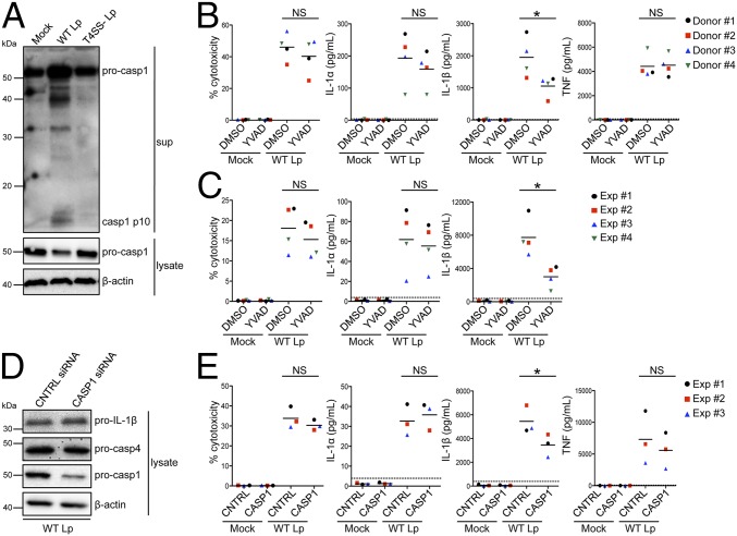
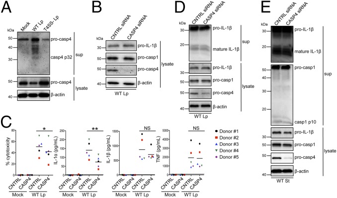
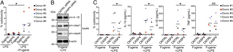
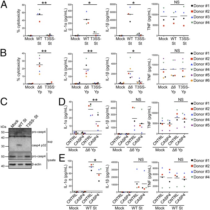
Inflammasomes are critical for host defense against bacterial pathogens. In murine macrophages infected by gram-negative bacteria, the canonical inflammasome activates caspase-1 to mediate pyroptotic cell death and release of IL-1 family cytokines. Additionally, a noncanonical inflammasome controlled by caspase-11 induces cell death and IL-1 release. However, humans do not encode caspase-11. Instead, humans encode two putative orthologs: caspase-4 and caspase-5. Whether either ortholog functions similar to caspase-11 is poorly defined. Therefore, we sought to define the inflammatory caspases in primary human macrophages that regulate inflammasome responses to gram-negative bacteria. We find that human macrophages activate inflammasomes specifically in response to diverse gram-negative bacterial pathogens that introduce bacterial products into the host cytosol using specialized secretion systems. In primary human macrophages, IL-1β secretion requires the caspase-1 inflammasome, whereas IL-1α release and cell death are caspase-1-independent. Instead, caspase-4 mediates IL-1α release and cell death. Our findings implicate human caspase-4 as a critical regulator of noncanonical inflammasome activation that initiates defense against bacterial pathogens in primary human macrophages.

Keywords: caspase-4; gram-negative bacteria; inflammasome; innate immunity; primary macrophages.


Conflict of interest statement

The authors declare no conflict of interest.

Figures

Fig. 1.
Fig. 1.
L. pneumophila induces both IL-1α and IL-1β release from human macrophages. Phorbol 12-myristate 13-acetate (PMA)-differentiated THP-1 cells (A and B) or primary human MDMs (C and D) were infected with WT Lp, infected with T4SS Lp, or mock-infected with PBS (Mock) for 20 h. (E and F) Primary human MDMs were primed with LPS and infected with WT Lp or T4SS Lp or mock-infected for 4 h. Cell death (% cytotoxicity) was measured using a lactate dehydrogenase release assay and normalized to mock-infected cells. IL-1α and IL-1β levels in the supernatants were measured by ELISA. Immunoblot analysis was performed on lysates for full-length IL-1β (pro–IL-1β), and blots were reprobed for β-actin as a loading control. Western blots (B, D, and F) are representative of at least three independent experiments. Shown are the pooled results of four independent experiments in THP-1 cells (A) or the pooled results of six independent infections of cells from different healthy human donors (C and E). Each data point shows the mean of triplicate infected wells. For A, *P < 0.05 and **P < 0.01 by unpaired t test. For C and E, ***P < 0.001, **P < 0.01, and *P < 0.05 by paired t test. The dashed line is the limit of detection.
Fig. 2.
Fig. 2.
Caspase-1–dependent and –independent inflammasomes are activated during infection of human macrophages. (A) Primary human MDMs were infected with WT Lp, infected with T4SS Lp, or mock-infected for 20 h. Immunoblot analysis was performed on supernatants (sup) for cleaved caspase-1 (casp1 p10) and on lysates for full-length caspase-1 (pro-casp1). Lysates were reprobed for β-actin. Primary human MDMs (B) or PMA-differentiated THP-1 cells (C) were pretreated with 40 μM caspase-1 inhibitor [Ac-YVAD-cmk (YVAD)] or vehicle control (DMSO) and infected with WT Lp or mock-infected for 20 h. (D and E) PMA-differentiated THP-1 cells were transfected with control siRNA (CNTRL) or siRNA against caspase-1 and infected with WT Lp or mock-infected for 20 h. Immunoblot analysis was performed on lysates for pro-casp1, pro-casp4, and pro–IL-1β, and blots were reprobed for β-actin. Cell death was measured using a lactate dehydrogenase release assay and normalized to mock-infected cells. IL-1α, IL-1β, and TNF levels in the supernatants were measured by ELISA. (A and D) Western blots are representative of three independent experiments. Shown are the pooled results of four independent infections of cells from different donors (B) or the pooled results of four (C) or three (E) independent experiments in THP-1 cells. Each data point shows the mean of triplicate infected wells. *P < 0.05 by paired t test (B and E) or unpaired t test (C). NS, not significant. The dashed line is the limit of detection.
Fig. 3.
Fig. 3.
Caspase-4 contributes to noncanonical inflammasome activation in L. pneumophila-infected primary human macrophages. (A) Primary human MDMs were infected with WT Lp, infected with T4SS Lp, or mock-infected for 20 h. Immunoblot analysis was performed on supernatants for cleaved caspase-4 (casp4 p32) and on lysates for pro-casp4. Lysates were reprobed for β-actin. Primary human MDMs (BD) or THP-1 cells (E) were transfected with control siRNA or siRNA against caspase-4 and infected with WT Lp or mock-infected for 20 h. Immunoblot analysis was performed on supernatants for cleaved IL-1β (mature IL-1β) and casp1 p10 and on lysates for pro–IL-1β, pro-casp1, and pro-casp4, and blots were reprobed for β-actin. Cell death was measured using a lactate dehydrogenase release assay and normalized to mock-infected cells. IL-1α, IL-1β, and TNF levels in the supernatants were measured by ELISA. Western blots (A, B, D, and E) are representative of at least three independent experiments. (C) Shown are the pooled results of five independent infections of cells from different donors. Each data point shows the mean of triplicate infected wells. **P < 0.01 and *P < 0.05 by paired t test. The dashed line is the limit of detection.
Fig. 4.
Fig. 4.
Caspase-4 mediates inflammasome activation in primary human macrophages in response to intracellular LPS. (A) Primary human MDMs were primed with Pam3CSK4 (Invivogen) and treated with extracellular LPS at the indicated concentrations, mock-transfected with Fugene HD (Promega) alone, or transfected with Fugene HD and LPS at the indicated concentrations for 20 h. (B and C) Primary human MDMs were transfected with control siRNA or siRNA against caspase-4, primed with Pam3CSK4, and mock-transfected with Fugene HD alone or transfected with Fugene HD and 2 μg/mL LPS for 20 h. Immunoblot analysis was performed on lysates for pro-casp1, pro-casp4, and pro–IL-1β, and blots were reprobed for β-actin. Cell death was measured using a lactate dehydrogenase release assay and normalized to LPS alone (A) or mock-transfected cells (C). IL-1α, IL-1β, and TNF levels in the supernatants were measured by ELISA. (B) Western blots are representative of at least four independent experiments. (A and C) Shown are the pooled results of five independent infections of cells from different donors. Each data point shows the mean of triplicate infected wells. *P < 0.05 by paired t test. The dashed line is the limit of detection.
Fig. 5.
Fig. 5.
Caspase-4 has a conserved role in noncanonical inflammasome activation against gram-negative bacterial pathogens. (A and C) Primary human MDMs were primed with LPS and infected with WT S. Typhimurium (WT St), infected with T3SS St, or mock-infected for 4 h. Immunoblot analysis was performed on supernatants for casp4 p32 and on lysates for pro-casp4. Blots were reprobed for β-actin. (B) Primary human MDMs were primed with LPS and infected with T3SS-expressing effectorless Y. pseudotuberculosis (Δ6 Yp) or T3SS Yp or mock-infected for 4 h. (D and E) Primary human MDMs were transfected with control siRNA or siRNA against caspase-4, primed with LPS, and infected with Δ6 Yp (D) or WT St (E) or mock-infected for 4 h. Cell death was measured using a lactate dehydrogenase release assay and normalized to mock-infected cells. IL-1α, IL-1β, and TNF levels in the supernatants were measured by ELISA. (C) Western blots are representative of at least three independent experiments. Shown are the pooled results of four (A and E), five (D), or six (B) independent infections of cells from different donors. Each data point shows the mean of triplicate infected wells. *P < 0.05 and **P < 0.01 by paired t test. The dashed line is the limit of detection.

References

    1. Janeway CA, Jr, Medzhitov R. Innate immune recognition. Annu Rev Immunol. 2002;20:197–216. - PubMed
    1. Harton JA, Linhoff MW, Zhang J, Ting JP-Y. Cutting edge: CATERPILLER: A large family of mammalian genes containing CARD, pyrin, nucleotide-binding, and leucine-rich repeat domains. J Immunol. 2002;169(8):4088–4093. - PubMed
    1. Vance RE, Isberg RR, Portnoy DA. Patterns of pathogenesis: Discrimination of pathogenic and nonpathogenic microbes by the innate immune system. Cell Host Microbe. 2009;6(1):10–21. - PMC - PubMed
    1. Martinon F, Burns K, Tschopp J. The inflammasome: A molecular platform triggering activation of inflammatory caspases and processing of proIL-beta. Mol Cell. 2002;10(2):417–426. - PubMed
    1. Li P, et al. Mice deficient in IL-1 beta-converting enzyme are defective in production of mature IL-1 beta and resistant to endotoxic shock. Cell. 1995;80(3):401–411. - PubMed

Publication types

MeSH terms