. 2015 Apr 27:9:91.
doi: 10.3389/fnbeh.2015.00091. eCollection 2015.

Differences in long-term memory stability and AmCREB level between forward and backward conditioned honeybees (Apis mellifera)

Affiliations

Differences in long-term memory stability and AmCREB level between forward and backward conditioned honeybees (Apis mellifera)

Johannes Felsenberg et al. Front Behav Neurosci. .

Abstract

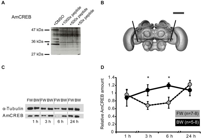
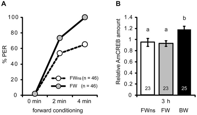
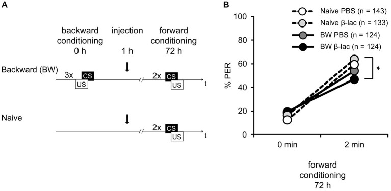
In classical conditioning a predictive relationship between a neutral stimulus (conditioned stimulus; CS) and a meaningful stimulus (unconditioned stimulus; US) is learned when the CS precedes the US. In backward conditioning the sequence of the stimuli is reversed. In this situation animals might learn that the CS signals the end or the absence of the US. In honeybees 30 min and 24 h following backward conditioning a memory for the excitatory and inhibitory properties of the CS could be retrieved, but it remains unclear whether a late long-term memory is formed that can be retrieved 72 h following backward conditioning. Here we examine this question by studying late long-term memory formation in forward and backward conditioning of the proboscis extension response (PER). We report a difference in the stability of memory formed upon forward and backward conditioning with the same number of conditioning trials. We demonstrate a transcription-dependent memory 72 h after forward conditioning but do not observe a 72 h memory after backward conditioning. Moreover we find that protein degradation is differentially involved in memory formation following these two conditioning protocols. We report differences in the level of a transcription factor, the cAMP response element binding protein (CREB) known to induce transcription underlying long-term memory formation, following forward and backward conditioning. Our results suggest that these alterations in CREB levels might be regulated by the proteasome. We propose that the differences observed are due to the sequence of stimulus presentation between forward and backward conditioning and not to differences in the strength of the association of both stimuli.

Keywords: CREB; backward conditioning; classical conditioning; long-term memory; proteasome; transcription; ubiquitin.


Figures

Figure 1
Figure 1
Late long-term memory is formed after forward conditioning but not after backward conditioning. (A) Two groups of honeybees were conditioned with three forward trials. Three hours after conditioning both groups were injected with either Act D or with the solvent PBS, 72 h after conditioning memory retention was tested. (B) The bees’ performance during the presentation of three forward conditioning trials does not differ between the two groups injected with either Act D (gray) or PBS (white) injection. The arrow indicates the time point of injection. Performance is lower in forward trained Act D-injected bees (gray) compared to PBS-injected bees (white) during the retention test at 72 h after conditioning. The asterisk indicates the significant differences (p < 0.05). (C) Retardation of acquisition assay. Honeybees were conditioned with three backward trials (backward) or were left untreated (Naive), 72 h after backward conditioning both groups received two forward conditioning trials. (D) The performance of bees during forward conditioning in the retardation of acquisition assay is not different between the Naive group (Naive, white) and the backward group (BW, gray).
Figure 2
Figure 2
The amount of AmCREB protein changes after conditioning. (A) Peptide competition assay to test the specificity of the anti-human CREB antibody (see methods) used in this study. Before incubating a western blot of honeybee brain homogenate with an anti-human CREB antibody the antibody was preincubated with a 1000x, 100x, and 50x molar excess of a peptide from the C-terminus of AmCREB. This peptide is homologous to the antibody’s human epitope. Bands disappearing following the AmCREB peptide preincubation are regarded as AmCREB proteins. The 33 kDa band (marked with a star) is analyzed in the subsequent experiments. Left: Size of the prestained protein marker. (B) The central part of the bee brain was analyzed (picture taken from the honeybee standard atlas, http://www.neurobiologie.fu-berlin.de/beebrain/DownloadGeneral.html) (Brandt et al., 2005). Black lines delimit the dissected part. Scale bar ~300 μm (gray line). (C,D) Two groups of honeybees were conditioned with either three forward trials (FW, gray) or three backward trials (BW, black). The forward conditioned animals were selected according to a conditioned response (CR) in the third trial. The brains were dissected at 1 h, 3 h, 6 h and 24 h after conditioning and probed for their relative amount of AmCREB. (C) Representative western blot for the FW and BW at the respective time points. Following protein separation and blotting, the membrane was cut at approximately 50 kDa horizontally in two. The upper part (>50 kDa) was probed with the anti-α-tubulin antibody and the lower part (<50 kDa) with the anti-CREB antibody. (D) Quantification of the relative amount of AmCREB shows a decreased AmCREB amount 3 h and 6 h after conditioning in the FW group compared to the BW group. The asterisks indicate the significant differences (p < 0.05).
Figure 3
Figure 3
The AmCREB amount depends on the timing of stimulus presentations during conditioning. (A) Honeybees were forward conditioned with three trials. The forward conditioned animals were divided into two groups: one was selected according to a CR in the third trial (FW, gray) and the other remained unselected (FWns, white). A third group received three backward trials (BW, trials not shown). (B) The brains were dissected 3 h after conditioning and analyzed for their relative AmCREB level. The quantification of the relative amount of AmCREB present shows a decreased amount in both forward conditioned groups compared to the backward conditioned bees. One sample consisted of two pooled brains. The numbers in the bars represent the sample size. The groups with unequal letters (a-b) differ significantly. The P-value was corrected due to multiple testing (p < 0.01). Whiskers represent the standard error.
Figure 4
Figure 4
Proteasome inhibition by β-lactone enhances late long-term memories (lLTM) upon forward conditioning. (A) The proteasomal targeting motif of human CREB (Taylor et al., 2000) matches the amino acid sequence of AmCREB. (B) The comparison of the amino acid sequence of monoubiquitin derived from the human precursor UBC protein (Catic and Ploegh, 2005) compared to the predicted Apis mellifera ubiquitin revealed that these sequences are highly homologous. Only one out of 76 amino acids is different (gray box). The sequences that are critical for polyubiquitination (K48; bold, C-terminus) are identical (underlined in black). (C) Western blot analysis of mono- and poly-ubiquitin in bee brain lysate. A slot of a SDS-PAGE gel was loaded with bee brain homogenate. Following protein separation and blotting, the membrane was cut vertically from top to bottom in the middle of the lane and probed with antibodies detecting K48-linked poly-ubiquitin or mono-ubiquitin. The analysis shows that the monoubiquitin antibody detects a single band at ~10 kDa, whereas the antibody against K48-linked polyubiquitin does not detect this band. Both antibodies detect a smear. (D–F) Honeybees were injected with 1 mM of β-lactone (β-lac, gray) or the solvent PBS (white). The brains were dissected at 5 min, 30 min and 60 min after injection. (D) In order to examine the level of polyubiquitination, the membranes were cut into two on the level of the 70 kDa marker band. The upper part was probed with the antibody detecting K48-linked poly-ubiquitin and the lower part with the anti-α-tubulin antibody. Quantification shows that the signal is increased 5 min after injection in the β-lac groups compared to the PBS control group. (E) Honeybees were conditioned with three forward trials or three backward trials. One hour after conditioning, the honeybees were injected with either 1 mM of β-lactone (β-lac) or with the solvent PBS. Three days after conditioning, their memory was tested with one CS only trial. (F) The performance during the three forward conditioning trials did not differ in the bees subjected to the injection one hour later (arrow). Forward trained animals injected with β-lac (light gray) show increased performance during the memory test at 72 h after conditioning compared to the PBS control group (white). The asterisk indicates significant differences (p < 0.05).
Figure 5
Figure 5
Proteasome activity does not gate lLTM for the CS excitatory and inhibitory properties following backward conditioning. (A) Honeybees were conditioned with three backward trials (BW) or remained naive (Naive). Three hours after conditioning, both groups were injected with either 1 mM of β-lactone (β-lac) or with the solvent PBS. Three days after conditioning all groups received two forward trials. (B) The performance of bees during the retardation test shows that backward conditioned bees injected with PBS (Naive PBS, white and BW PBS, dark gray) were not different from each other and from the respective β-lac groups, indicating that no memory for the CS inhibitory properties was formed. However, β-lac-injected bees (BW β-lac, black) show a reduced response in the second acquisition trial compared to the Naive β-lac-injected bees (light gray) suggesting that proteasome activity to a small extent impacts the acquisition of excitatory AND inhibitory properties. The asterisk indicates significant differences of the post hoc test (p < 0.05).

Similar articles

Cited by

References

    1. Alberini C. M. (2009). Transcription factors in long-term memory and synaptic plasticity. Physiol. Rev. 89, 121–145. 10.1152/physrev.00017.2008 - DOI - PMC - PubMed
    1. Barot S. K., Kyono Y., Clark E. W., Bernstein I. L. (2008). Visualizing stimulus convergence in amygdala neurons during associative learning. Proc. Natl. Acad. Sci. U S A 105, 20959–20963. 10.1073/pnas.0808996106 - DOI - PMC - PubMed
    1. Boto T., Louis T., Jindachomthong K., Jalink K., Tomchik S. M. (2014). Dopaminergic modulation of cAMP drives nonlinear plasticity across the Drosophila mushroom body lobes. Curr. Biol. 24, 822–831. 10.1016/j.cub.2014.03.021 - DOI - PMC - PubMed
    1. Brandt R., Rohlfing T., Rybak J., Krofczik S., Maye A., Westerhoff M., et al. . (2005). Three-dimensional average-shape atlas of the honeybee brain and its applications. J. Comp. Neurol. 492, 1–19. 10.1002/cne.20644 - DOI - PubMed
    1. Catic A., Ploegh H. L. (2005). Ubiquitin–conserved protein or selfish gene? Trends Biochem. Sci. 30, 600–604. 10.1016/j.tibs.2005.09.002 - DOI - PubMed

LinkOut - more resources