. 2015 May 12;11(5):e1004895.
doi: 10.1371/journal.ppat.1004895. eCollection 2015 May.

Edin Expression in the Fat Body Is Required in the Defense Against Parasitic Wasps in Drosophila melanogaster

Affiliations

Edin Expression in the Fat Body Is Required in the Defense Against Parasitic Wasps in Drosophila melanogaster

Leena-Maija Vanha-Aho et al. PLoS Pathog. .

Abstract

The cellular immune response against parasitoid wasps in Drosophila involves the activation, mobilization, proliferation and differentiation of different blood cell types. Here, we have assessed the role of Edin (elevated during infection) in the immune response against the parasitoid wasp Leptopilina boulardi in Drosophila melanogaster larvae. The expression of edin was induced within hours after a wasp infection in larval fat bodies. Using tissue-specific RNAi, we show that Edin is an important determinant of the encapsulation response. Although edin expression in the fat body was required for the larvae to mount a normal encapsulation response, it was dispensable in hemocytes. Edin expression in the fat body was not required for lamellocyte differentiation, but it was needed for the increase in plasmatocyte numbers and for the release of sessile hemocytes into the hemolymph. We conclude that edin expression in the fat body affects the outcome of a wasp infection by regulating the increase of plasmatocyte numbers and the mobilization of sessile hemocytes in Drosophila larvae.


Conflict of interest statement

The authors have declared that no competing interests exist.

Figures

Fig 1
Fig 1. Edin expression is induced upon a wasp infection.
(A) Wasp infection causes a 6.7-fold increase in edin expression in 2nd instar Canton S larvae. Data are pooled from two independent experiments, n = 2 for each experiment, where one sample represents 10 larvae. (B) Edin expression is induced in the fat bodies of Canton S larvae 24 hours post infection. The data are pooled from four independent experiments, and each experiment consisted of two samples, where one sample represents 8–10 larval fat bodies.
Fig 2
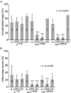
Fig 2. Knock down of edin in the fat body decreases the encapsulation and killing ability of Drosophila larvae.
(A) The encapsulation response of two different edin RNAi lines (edin 14289 and edin 109528) was analyzed 27-29h after a wasp infection. The C564-GAL4 (C564>), Fb-GAL4 (Fb>) and Hml Δ;He-GAL4 (HH>) drivers were used to drive the expression of the RNAi constructs. w 1118 (w) was used as control. Data were pooled from one to eight individual experiments, as depicted on each column, each experiment with at least 50 analyzed individual infected larvae. (B) The ability of Drosophila larvae to kill wasp eggs was assessed with two different edin RNAi lines (edin 14289 and edin 109528) 48-50h after infection. The C564-GAL4 (C564>) and Fb-GAL4 (Fb>) drivers were used to drive the expression of the RNAi constructs. w 1118 (w) was used as control. Data are pooled from three to sixteen independent experiments, as indicated on each column, and at least 50 infected larvae were scored per experiment. Error bars in A and B show standard deviations. Knocking down the expression of edin in several tissues including the fat body or in the fat body alone caused a significant decrease in the encapsulation activity and killing response of Drosophila larvae compared to controls, whereas knocking down edin in hemocytes had no effect.
Fig 3
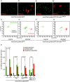
Fig 3. Quantification of hemocytes in edin RNAi larvae after a wasp infection.
(A-B) Hemocytes of infected larvae were bled 48–50 hours post-infection and visualized with the eaterGFP (green) and msnCherry (red) reporters. Uninfected controls contained only GFP-positive cells that corresponded to plasmatocytes (green). (A’ and B’) msnCherry expression was detected in the infected samples and this included lamellocytes (asterisks) and cells that express both eaterGFP and msnCherry indicating that they were undergoing lamellocyte transition. Lamellocytes were present also in the infected edin RNAi larvae suggesting that edin expression is not necessary for lamellocyte differentiation. Scale bars are 10 μm (C-E) Flow cytometry was carried out to quantify the amount of hemocytes in the unchallenged and the wasp infected edin RNAi larvae. (C = control, inf = infected)
Fig 4
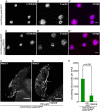
Fig 4. Edin expression in fat body is dispensable for normal hemocyte attachment to and spreading on glass and wasp eggs, but is necessary to increase blood cell numbers in circulation early after wasp infection.
(A-B”) Hemocytes from infected control larvae (msnCherry,eaterGFP, A-A”) and from infected larvae in which edin was knocked down in the fat body (msnCherry,eaterGFP;Fb>edin 109528, B-B”) spread normally on glass 14 hours after wasp infection despite knock down of edin in fat body. The spreading ability of hemocytes was assayed by staining α-Tubulin (blue) and F-actin (magenta). The size bar denotes 10 μm. (C and D). Wasp eggs from infected control larvae (msnCherry,eaterGFP, C) and from infected larvae in which edin was knocked down in the fat body (msnCherry,eaterGFP;Fb>edin 109528, D) were stained with the anti-plasmatocyte antibody NimC1. The wasp eggs were dissected 14 hours after parasitization and are still attached to the gut. Plasmatocytes spread normally on the eggs irrespective of edin RNAi in the fat body. Arrows denote examples of plasmatocytes spreading and adhering normally on the surface of the wasp egg. The scale bar depicts 50 μm. (E) Edin RNAi in the fat body (msnCherry,eaterGFP;Fb>edin 109528) reduced the number of circulating cells after wasp infection in comparison to control larvae (msnCherry,eaterGFP) 14 hours after infection. Circulating blood cell numbers were obtained with flow cytometry.
Fig 5
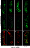
Fig 5. Edin expression in the fat body is required for the activation of plasmatocytes upon a wasp attack 27–29 hours after infection.
The in vivo phenotype of wasp infected edin RNAi larvae was studied using the eaterGFP (green = plasmatocytes) and mCherry (red = lamellocytes) reporters. Imaging was performed 27–29 hours post infection with living Drosophila larvae. (A-D) Uninfected larvae show an uninterrupted banding pattern formed by sessile plasmatocytes (green). (E-H) Shows only the green channel (eaterGFP) of infected larvae and (E’-H’) both the green and the red (msnCherry) channel. Infected larvae have lost the banding pattern and lamellocytes have appeared, but infected msnCherry,eaterGFP;Fb>edin 109528 larvae still show a visible banding pattern formed by the sessile cells. = uninfected larvae, + = wasp infected larvae. Fig 5 shows representative images of at least 10 larvae per condition and per genotype.
Fig 6
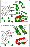
Fig 6. A schematic presentation of the function of Edin.
(1.) Edin is induced in the fat body shortly after wasp infection and secreted into the hemolymph. There, Edin directly or indirectly induces the release of plasmatocytes from the sessile hemocyte compartment. These cells go into circulation, find the wasp egg and participate in forming the capsule around the parasitoid egg. (2.) If the expression of edin is knocked down in the fat body in the context of a wasp infection, plasmatocytes are retained in the sessile compartment instead of being released into circulation, causing a defect in the encapsulation of the wasp egg.

Similar articles

Cited by

References

    1. Keebaugh ES, Schlenke TA. Insights from natural host-parasite interactions: The drosophila model. Dev Comp Immunol 2014; 42(1): 111–123. 10.1016/j.dci.2013.06.001 - DOI - PMC - PubMed
    1. Benassi V, Coustau C, Carton Y. Insect immunity: A genetic factor (hrtp) is essential for antibacterial peptide expression in drosophila after infection by parasitoid wasps. Arch Insect Biochem Physiol 2000; 43(2): 64–71. - PubMed
    1. Schlenke TA, Morales J, Govind S, Clark AG. Contrasting infection strategies in generalist and specialist wasp parasitoids of drosophila melanogaster. PLoS Pathog 2007; 3(10): 1486–1501. - PMC - PubMed
    1. Wertheim B, Kraaijeveld AR, Schuster E, Blanc E, Hopkins M, et al. Genome-wide gene expression in response to parasitoid attack in drosophila. Genome Biol 2005; 6(11): R94 - PMC - PubMed
    1. Valanne S, Wang JH, Rämet M. The drosophila toll signaling pathway. J Immunol 2011; 186(2): 649–656. 10.4049/jimmunol.1002302 - DOI - PubMed

Publication types

MeSH terms