. 2015 May 12;10(5):e0127039.
doi: 10.1371/journal.pone.0127039. eCollection 2015.

Optimization of translation profiles enhances protein expression and solubility

Affiliations

Optimization of translation profiles enhances protein expression and solubility

Anne-Katrin Hess et al. PLoS One. .

Abstract

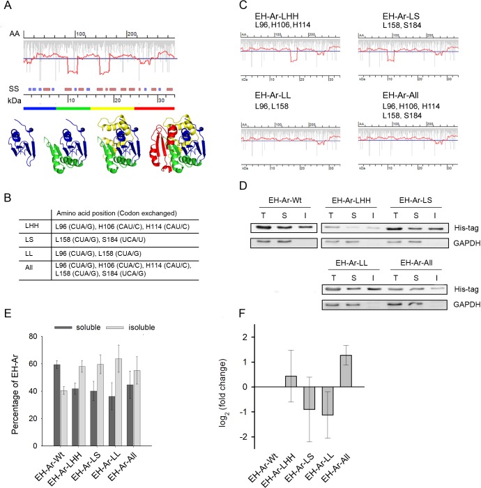
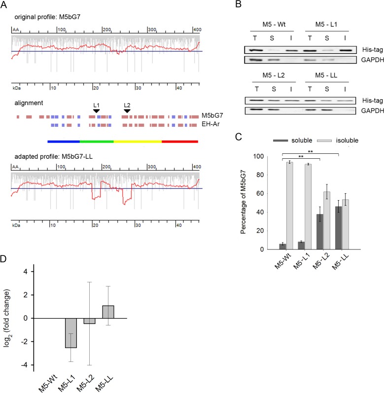
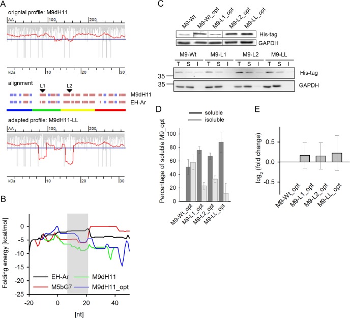
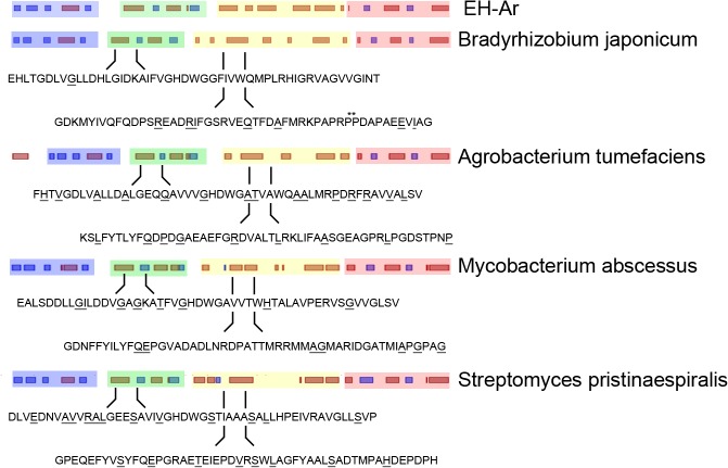
mRNA is translated with a non-uniform speed that actively coordinates co-translational folding of protein domains. Using structure-based homology we identified the structural domains in epoxide hydrolases (EHs) and introduced slow-translating codons to delineate the translation of single domains. These changes in translation speed dramatically improved the solubility of two EHs of metagenomic origin in Escherichia coli. Conversely, the importance of transient attenuation for the folding, and consequently solubility, of EH was evidenced with a member of the EH family from Agrobacterium radiobacter, which partitions in the soluble fraction when expressed in E. coli. Synonymous substitutions of codons shaping the slow-transiting regions to fast-translating codons render this protein insoluble. Furthermore, we show that low protein yield can be enhanced by decreasing the free folding energy of the initial 5'-coding region, which can disrupt mRNA secondary structure and enhance ribosomal loading. This study provides direct experimental evidence that mRNA is not a mere messenger for translation of codons into amino acids but bears an additional layer of information for folding, solubility and expression level of the encoded protein. Furthermore, it provides a general frame on how to modulate and fine-tune gene expression of a target protein.


Conflict of interest statement

Competing Interests: The authors have declared that no competing interests exist.

Figures

Fig 1
Fig 1. Translation attenuation sites delineate domain boundaries and impact protein solubility.
(A) Translation profile of EH-Ar predicted with RiboTempo. Vertical gray bars represent the rate of translation of each single codon which is averaged (red line) along the whole ORF with a window of 19 codons [42]. Translation minima below the genome-wide threshold (blue horizontal line) denote the putative slow-translating attenuation sites. AA denotes amino acid number, kDa marks the corresponding molecular weight and SS denotes the predicted secondary structure (β-sheets—blue bars, α-helices—dark red bars, uncolored empty space—linking structural elements). The rainbow-colored bar visualizes the putative structural domains, colored in the same way in the 3D- structure (PDB 1EHY). (B) Summary of the exchanged codons in EH-Ar. The position of the exchanged amino acids for each variant is indicated. (C) Translation profiles of EH-Ar variants with exchanged slow translating patches (B) predicted with RiboTempo. (D, E). Removal of the translational attenuation sites reduces the solubility of EH-Ar. (D) Representative immunoblot of EH-Ar variants (summarized in C). The total (T) protein content was fractionated into soluble (S) and insoluble (I) fractions and 0.05 OD600 of cells were applied per lane. GAPDH served as a loading control; note that it is a completely soluble protein and its absence in the insoluble fraction confirms the good quality of the fractionation procedure. (E) Quantification of the immunoblots of three biological replicates ± SEM. Each total fraction was normalized to GAPDH intensity to allow for comparison between the samples; the soluble and insoluble fractions were determined as a percentage of this normalized value. *, p<0.05, Tukey’s test. (F) Quantification of mRNA levels by qRT-PCR of the EH-Ar variants. Values were normalized to GAPDH mRNA expression, represented as a fold-change to the wild-type mRNA and are means ± SEM (n = 3).
Fig 2
Fig 2. Introducing translational attenuation sites into the EH M5bG7 sequence enhances its solubility.
(A) Translation profile of wild-type M5bG7 (top diagram, original profile) and upon introduction of translational attenuation sites (bottom diagram, adapted profile) in E. coli. Secondary structure alignment (β-sheets—blue bars, α-helices—dark red bars, uncolored empty space—linking structural elements) of M5bG7 to EH-Ar to identify regions for introduction of slow-translating stretches (indicated with arrows). The domains in M5bG7 were delineated based on the domain architecture of EH-Ar represented in the color code as in Fig 1A. Note the longer N-terminal domain of M5bG7 than that of EH-Ar. (B) Representative immunoblot of M5bG7 (abbreviated M5) variants. L1 (Leu 189, CUC/A, numbering is according to the M5bG7 sequence) and L2 (Leu 257, CUG/A) denote the synonymous exchange of a fast-translating Leu codons to Leu CUA in the first and second attenuation, respectively, and LL (Leu189, CUC/A, Leu257, CUG/A) in both simultaneously. T, total protein, S, soluble and I, insoluble fraction. GAPDH served as loading control. (C) Quantification of immunoblots of three biological replicates ± SEM. **, p<0.01, Tukey’s test. For details refer to the legend of Fig 1. (D) Quantification of mRNA levels by qRT-PCR of the M5bG7 variants. Values were normalized to GAPDH mRNA expression, represented as a fold-change to the wild-type mRNA and are means ± SEM (n = 3).
Fig 3
Fig 3. Introducing translational attenuation sites into the EH M9dH11 sequence enhances its solubility.
(A) Translation profile of wild-type M9dH11 (top diagram, original profile) and upon introduction of translational attenuation sites (bottom diagram) in E. coli. Secondary structure alignment (β-sheets—blue bars, α-helices—dark red bars, uncolored empty space—linking structural elements) of M9dH11 to EH-Ar to identify regions for introduction of slow-translating stretches (indicated with arrows). The domains in M9dH11 were delineated based on the EH-Ar domain architecture presented in the color code used in Fig 1A. (B) Local folding energies of the mRNA sequences of EH variants around the start codon. The zero nucleotide position corresponds to first nucleotide of the start ATG codon. The shadowed area marks the codons (3rd to 7th), at which changes were undertaken in M9dH11_opt. The -50 nucleotides for all four sequences are identical and determined from the upstream region of the expression vector. For comparison, two EH variants, EH-Ar and M5bG7, with lower folding energies than that of M9dH11 are included. The energy of M9dH11_opt falls between that of EH-Ar and M5bG7. (C) Representative immunoblot (n = 3) of total expression (upper panel) of M9dH11 (abbreviated M9) variants and fractionation to soluble and insoluble fractions (bottom panel). M9_opt denotes the variants with optimized secondary structure at the 5’-end. L1 (Leu 88, CUG/A, numbering is according to the M9dH11 sequence), L2 (Leu154, CUG/A) and LL (Leu88, CUG/A, Leu154, CUG/A) denote synonymous exchange of a fast-translating Leu codon to Leu CUA in the first, second or both attenuation sites (LL), respectively. T, total protein, S, soluble and I, insoluble fraction. GAPDH served as loading control. For details refer to the legend to Fig 1. (D) Quantification of immunoblots of two biological replicates ± SD. (E) Quantification of mRNA levels by qRT-PCR of the M9dH11_opt variants. Values were normalized to GAPDH mRNA expression, represented as a fold-change to the wild-type mRNA and are means ± SEM (n = 3).
Fig 4
Fig 4. Overview of EHs from different species.
Secondary structure alignment (β-sheets—blue bars, α-helices—dark red bars, uncolored empty space—linking structural elements) of EHs from different organisms [54]. The domains are color coded according to EH-Ar crystal structure (PDB 1EHY, Fig 1A). Downstream of the domain boundaries (20–70 aa), according to the identified slow-translating regions of EH-Ar (translation profile, Fig 1A), the amino acid sequences of each EH is shown and the amino acids encoded by rare codons are underlined. Rare codons are defined as the ten least used codons for each species.

Similar articles

Cited by

References

    1. Komili S, Silver PA. Coupling and coordination in gene expression processes: a systems biology view. Nat Rev Genet. 2008;9: 38–48. - PubMed
    1. Turner M. Is transcription the dominant force during dynamic changes in gene expression? Adv Exp Med Biol. 2011;780: 1–13. 10.1007/978-1-4419-5632-3_1 - DOI - PubMed
    1. Rouskin S, Zubradt M, Washietl S, Kellis M, Weissman JS. Genome-wide probing of RNA structure reveals active unfolding of mRNA structures in vivo. Nature. 2014;505: 701–5. 10.1038/nature12894 - DOI - PMC - PubMed
    1. Bentele K, Saffert P, Rauscher R, Ignatova Z, Blüthgen N. Efficient translation initiation dictates codon usage at gene start. Mol Syst Biol. 2013;9: 675 10.1038/msb.2013.32 - DOI - PMC - PubMed
    1. Goodman DB, Church GM, Kosuri S. Causes and effects of N-terminal codon bias in bacterial genes. Science. 2013;342: 475–9. 10.1126/science.1241934 - DOI - PubMed

MeSH terms

LinkOut - more resources