. 2015 May 12;10(5):e0125836.
doi: 10.1371/journal.pone.0125836. eCollection 2015.

Treatment with Vitamin D/MOG Association Suppresses Experimental Autoimmune Encephalomyelitis

Affiliations

Treatment with Vitamin D/MOG Association Suppresses Experimental Autoimmune Encephalomyelitis

Fernanda Chiuso-Minicucci et al. PLoS One. .

Erratum in

Abstract

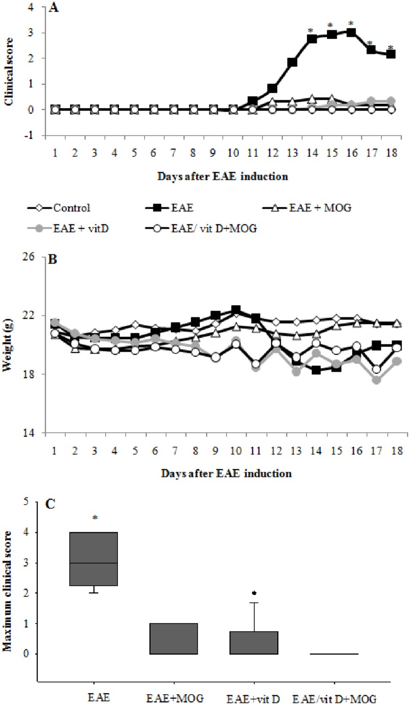
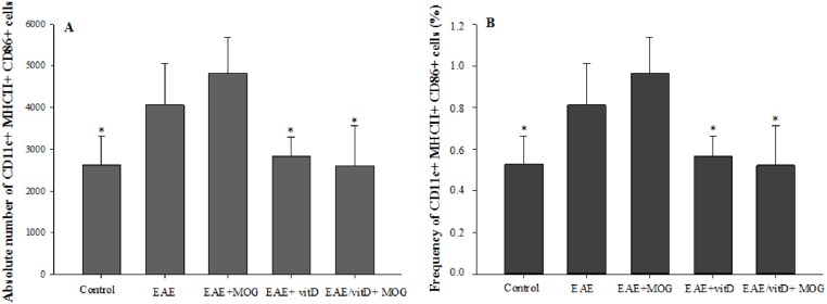
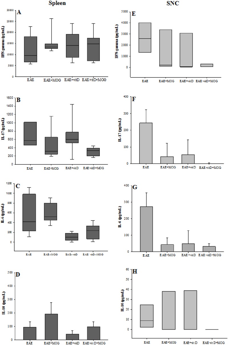
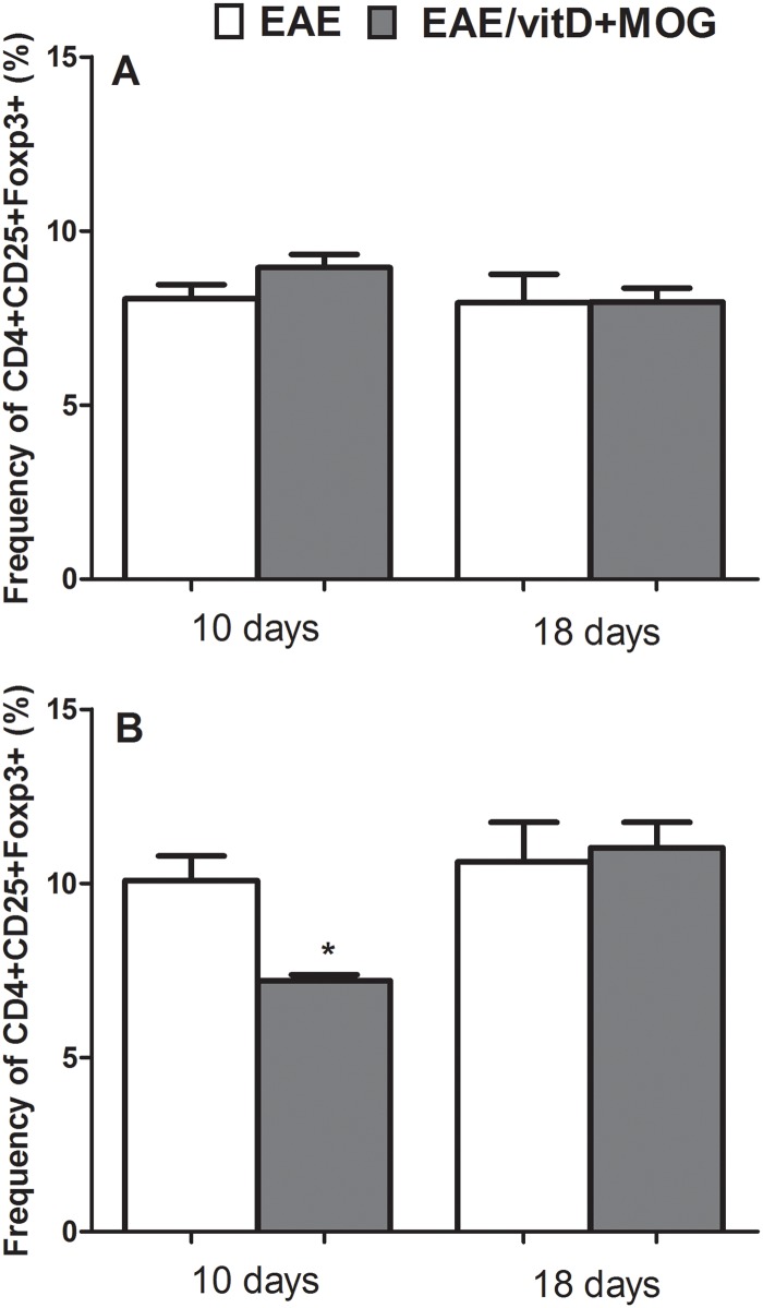
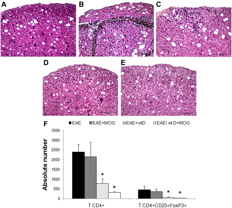
Experimental autoimmune encephalomyelitis (EAE) is an animal model to study multiple sclerosis (MS). Considering the tolerogenic effects of active vitamin D, we evaluated the therapeutic effect of myelin oligodendrocyte glycoprotein (MOG) associated with active vitamin D in EAE development. EAE was induced in female C57BL/6 mice by immunization with MOG emulsified with Complete Freund's Adjuvant plus Mycobacterium tuberculosis. Animals also received two intraperitoneal doses of Bordetella pertussis toxin. One day after immunization, mice were treated with 0,1 μg of 1α,25-dihydroxyvitamin D3 (1,25(OH)2D3) every other day during 15 days (on days 1, 3, 5, 7, 9, 11, 13 and 15). MOG (150 μg) was co-administered on days 3 and 11. The administration of 1,25(OH)2D3 or MOG determined significant reduction in EAE incidence and in clinical scores. When MOG was associated with 1,25(OH)2D3 the animals did not develop EAE. Spleen and central nervous system (CNS) cell cultures from this group produced less IL-6 and IL-17 upon stimulation with MOG in comparison to the EAE control group. In addition, this treatment inhibited dendritic cells maturation in the spleen and reduced inflammatory infiltration in the CNS. The association of MOG with 1,25(OH)2D3 was able to control EAE development.


Conflict of interest statement

Competing Interests: The authors have declared that no competing interests exist.

Figures

Fig 1
Fig 1. Effect of treatment with 1,25(OH)2D3+MOG association on EAE development.
Kinetics of clinical scores (A), maximum clinical score (B) and weight variation (C). Comparisons between groups were made by one way ANOVA followed by Tukey’s test for parametric variables (A and C) and by Kruskal-Wallis followed by Dunn’s test for non-parametric variables (B). Results were expressed as mean or medians (25–75% ranges) of 12 animals per group. * p<0.05 compared with EAE+MOG, EAE+vitD and EAE/vitD+MOG groups. Data are representative of two independent experiments.
Fig 2
Fig 2. Quantification of splenic dendritic cells in C57BL/6 mice treated with 1,25(OH)2D3+MOG.
Absolute number (A) and percentage (B) of CD11c+MHCII+CD86+ cells in 500,000 acquired events. Comparisons between groups were made by one way ANOVA followed by Tukey’s test. Results are expressed as mean ± SD of 5 animals per group. * p<0.05 compared with EAE and EAE+MOG group. Data are representative of two independent experiments.
Fig 3
Fig 3. Cytokine production by spleen and CNS cell cultures.
IFN-γ (A and E), IL-17 (B and F), IL-6 (C and G) and IL-10 (D and H) levels were measured in spleen and CNS cell cultures stimulated with MOG. Comparisons between groups were made by one way ANOVA followed by Tukey’s test for parametric variables (D, F and G) and by Kruskal-Wallis followed by Dunn’s test for non-parametric variables (A, B, C, E and H). Data were presented by mean ± SE or medians (25–75% ranges) of 9 animals per group in spleen cultures or 4 pools (each pool contains cells from brain and spinal cord of 3 mice) per group in CNS cultures. * p<0.05. Data are representative of two independent experiments.
Fig 4
Fig 4. Quantification of regulatory T cells in peripheral lymphoid organs from C57BL/6 mice treated with 1,25(OH)2D3+MOG.
Percentage of CD4+CD25+Foxp3+ T cells in total CD4+ T cell. Spleen (A) and lymph nodes (B) were collected 10 and 18 days after EAE induction. Analyses were performed in 100,000 acquired events. Comparisons between groups were made by t-test followed by Tukey’s test for parametric variables. Results were expressed as mean of 6–9 animals per group. Data are representative of two independent experiments.
Fig 5
Fig 5. Infiltrating cells in the CNS after treatment of 1,25(OH)2D3+MOG.
Inflammatory process in lumbar spinal cord sections stained with H&E in control (A), EAE (B), EAE+MOG (C), EAE+vitD (D) and EAE/vitD+MOG (E) groups and absolute number of CD4+ T cells and CD4+CD25+Foxp3+ T cells in CNS-mononuclear cells (F). Regulatory T cells were evaluated in the total CD4+ T cell population and analyses were performed in 20,000 acquired events. Comparisons between groups were made by one way ANOVA followed by Tukey’s test. Data were presented by mean ± SE of 4 pools (each pool contains cells from brain and spinal cord of 3 mice) per group in CNS cultures cells. * p<0.05 in compared with EAE and EAE+MOG group. Scale bar = 100μm. Micrographs are representative of 5 animals per group. Data are representative of two independent experiments.
Fig 6
Fig 6. Effect of 1,25(OH)2D3 (alone or combined with MOG) treatment in calcium and phosphorus levels.
Serum calcium (A) and phosphorus (B) levels were quantified 18 days after EAE induction. Comparisons between groups were made by one way ANOVA followed by Tukey’s test. Data were presented by mean ± SE of 4–6 animals per group. * p<0.05 compared with Control, EAE, and EAE+MOG group. Data are representative of two independent experiments.

References

    1. Loleit V, Biberacher V, Hemmer B. Current and future therapies targeting the immune system in multiple sclerosis. Curr Pharm Biotechnol. 2014;15: 276–296. - PubMed
    1. Fletcher JM, Lalor SJ, Sweeney CM, Tubridy N, Mills KH. T cells in multiple sclerosis and experimental autoimmune encephalomyelitis. Clin Exp Immunol. 2010;162: 1–11. 10.1111/j.1365-2249.2010.04143.x - DOI - PMC - PubMed
    1. Mitchell KM, Dotson AL, Cool KM, Chakrabarty A, Benedict SH, LeVine SM. Deferiprone, an orally deliverable iron chelator, ameliorates experimental autoimmune encephalomyelitis. Mult Scler. 2007;13: 1118–1126. - PubMed
    1. Domingues HS, Mues M, Lassmann H, Wekerle H, Krishnamoorthy G. Functional and pathogenic differences of Th1 and Th17 cells in experimental autoimmune encephalomyelitis. PLoS One. 2010;5: e15531 10.1371/journal.pone.0015531 - DOI - PMC - PubMed
    1. Chen SJ, Wang YL, Fan HC, Lo WT, Wang CC, Sytwu HK. Current status of the immunomodulation and immunomediated therapeutic strategies for multiple sclerosis. Clin Dev Immunol. 2012;2012: 970789 10.1155/2012/970789 - DOI - PMC - PubMed

Publication types

MeSH terms