. 2015 May 12;10(5):e0126576.
doi: 10.1371/journal.pone.0126576. eCollection 2015.

SP-R210 (Myo18A) Isoforms as Intrinsic Modulators of Macrophage Priming and Activation

Affiliations

SP-R210 (Myo18A) Isoforms as Intrinsic Modulators of Macrophage Priming and Activation

Linlin Yang et al. PLoS One. .

Abstract

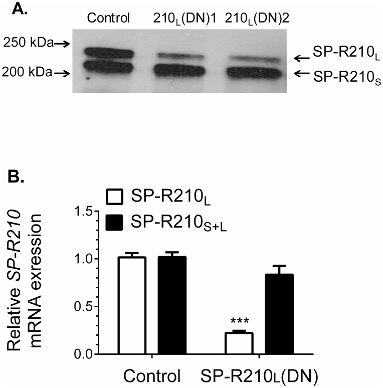
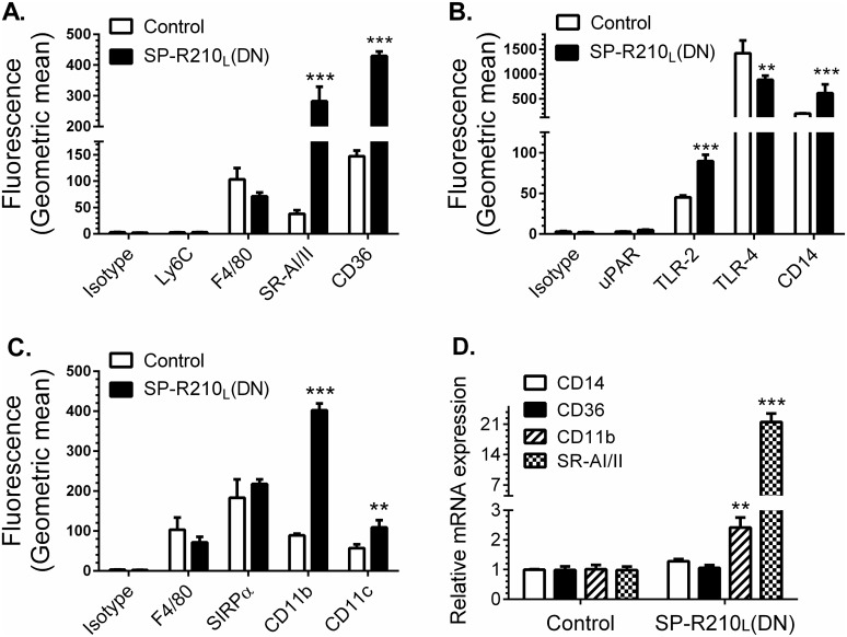
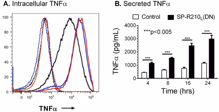
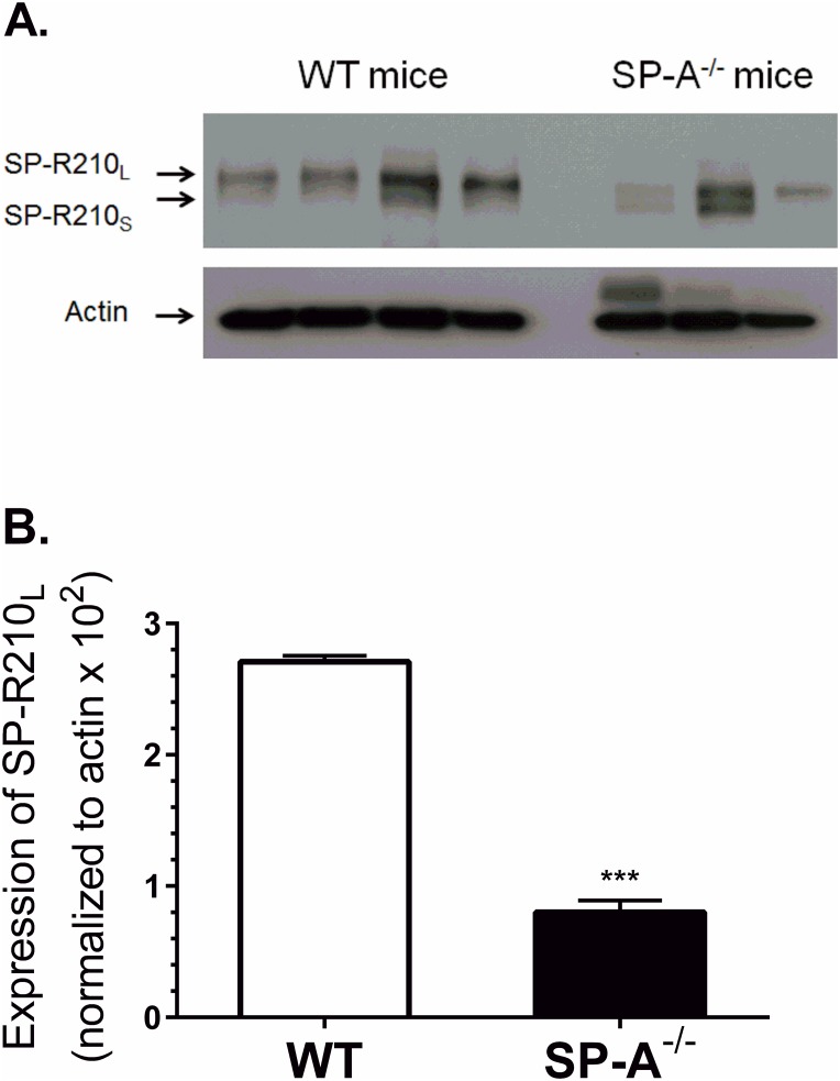
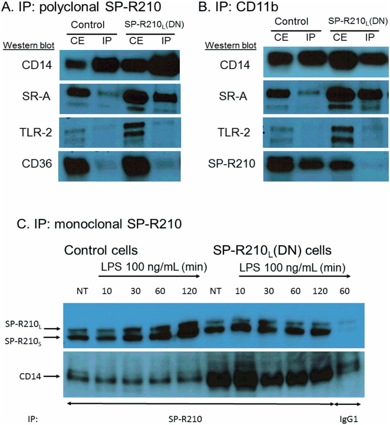
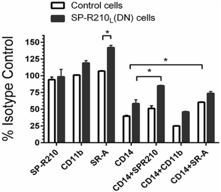
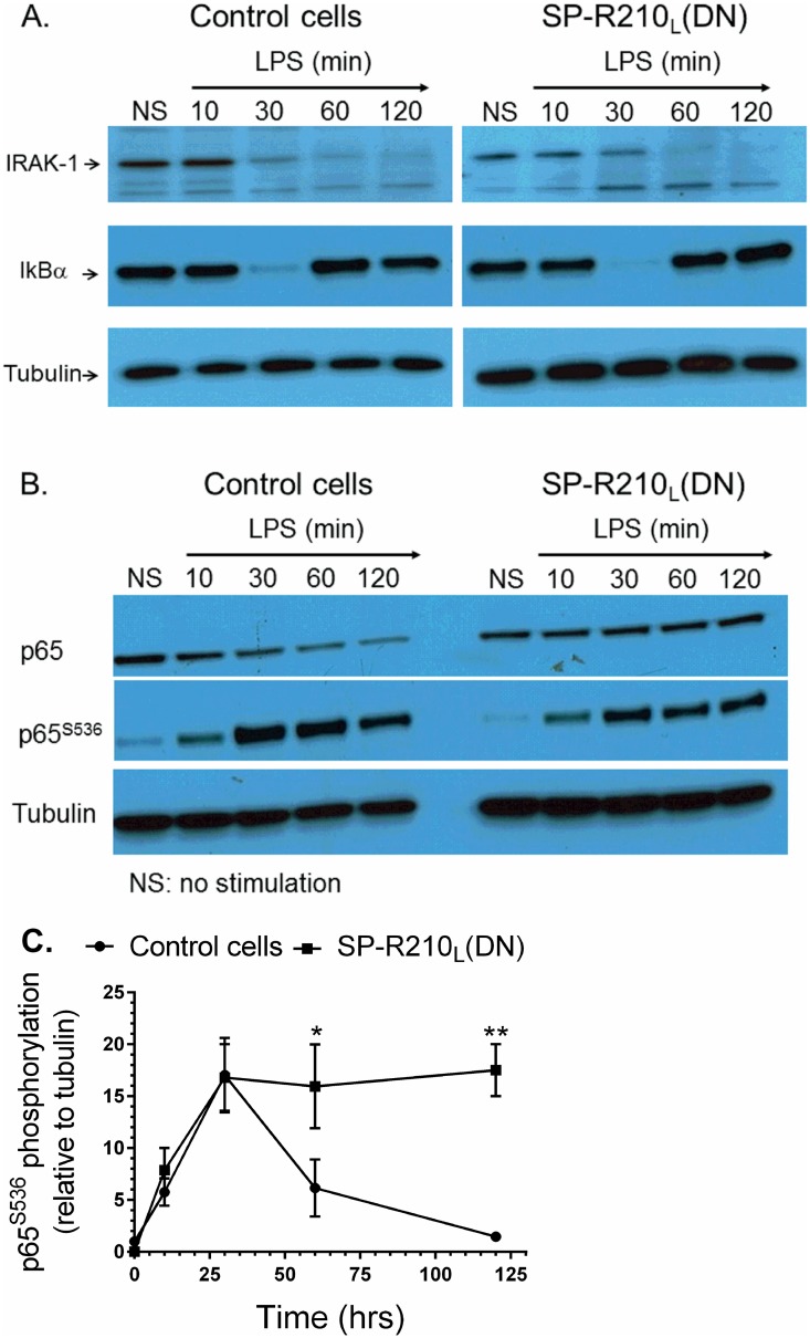
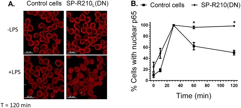
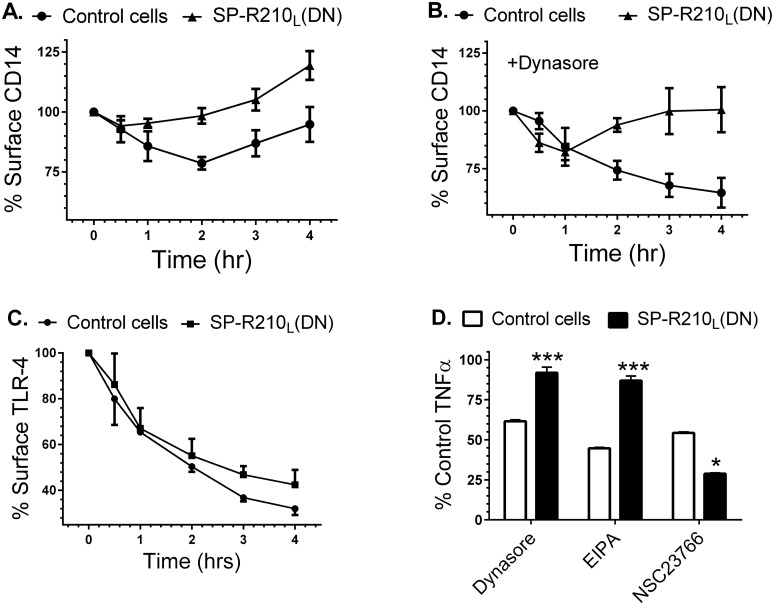
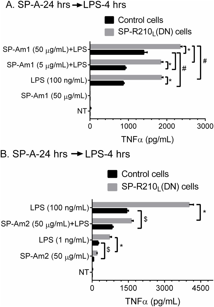
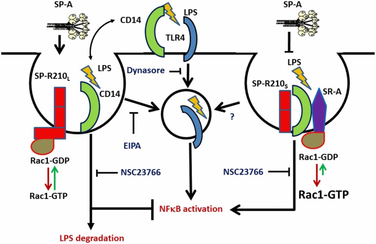
The surfactant protein (SP-A) receptor SP-R210 has been shown to increase phagocytosis of SP-A-bound pathogens and to modulate cytokine secretion by immune cells. SP-A plays an important role in pulmonary immunity by enhancing opsonization and clearance of pathogens and by modulating macrophage inflammatory responses. Alternative splicing of the Myo18A gene results in two isoforms: SP-R210S and SP-R210L, with the latter predominantly expressed in alveolar macrophages. In this study we show that SP-A is required for optimal expression of SP-R210L on alveolar macrophages. Interestingly, pre-treatment with SP-A prepared by different methods either enhances or suppresses responsiveness to LPS, possibly due to differential co-isolation of SP-B or other proteins. We also report that dominant negative disruption of SP-R210L augments expression of receptors including SR-A, CD14, and CD36, and enhances macrophages' inflammatory response to TLR stimulation. Finally, because SP-A is known to modulate CD14, we used a variety of techniques to investigate how SP-R210 mediates the effect of SP-A on CD14. These studies revealed a novel physical association between SP-R210S, CD14, and SR-A leading to an enhanced response to LPS, and found that SP-R210L and SP-R210S regulate internalization of CD14 via distinct macropinocytosis-like mechanisms. Together, our findings support a model in which SP-R210 isoforms differentially regulate trafficking, expression, and activation of innate immune receptors on macrophages.


Conflict of interest statement

Competing Interests: The authors have declared that no competing interests exist.

Figures

Fig 1
Fig 1. Dominant-negative disruption of SP-R210L.
Raw264.7 cells were stably transfected with empty pTriexNeo2 control or vector containing the 300 (210L(DN1)) and 350 (210L(DN2)) bp cDNA of SP-R210 carboxy-terminal isoforms (6). A) Detergent extracts were analyzed by Western blotting using affinity purified polyclonal antibodies recognizing both SP-R210L and SP-R210S. Lanes were loaded with 5 μg of protein. B) Total RNA from indicated cell lines was reverse transcribed and quantitated by qRT-PCR using TaqMan assays and primers encompassing the SP-R210L-specific exons 1 and 2 (red bars) and internal primers encompassing exon 17 and 18 common to both SP-R210 isoforms (black bars) and 18S rRNA as internal control. (n = 4 ***p<0.001).
Fig 2
Fig 2. Depletion of SP-R210L differentially enhances expression of innate immune receptors in macrophages.
Control and SP-R210L(DN) cells were analyzed by flow cytometry using indicated APC (A) or PE-conjugated antibodies (B, C) (n = 4–8). (D) mRNA levels of indicated receptors in SP-R210L(DN) cells relative to control cells were determined by qRT-PCR (n = 4 independent experiments performed in duplicate, **p<0.02, ***p<0.005).
Fig 3
Fig 3. Increased responsiveness of SP-R210L(DN) cells to LPS.
Control and SPR210L(DN) cells were plated in 12-well tissue culture dishes at a density of 150,000 cells/well and cultured 24 hrs in RPMI/10% FBS. Cells were then treated with 100 ng/mL LPS for 2, 4, 8, 16, and 24 hrs. A) Intracellular staining of TNFα was performed in brefeldin A blocked cells 2 hrs after treatment with LPS. Dotted histograms show untreated cells. Black, red, and blue histograms show intracellular TNFα staining of control, SP-R210L(DN)1, and SP-R210L(DN)2 cells. B) The levels of secreted TNFα were measured by ELISA in media at indicated time points after treatment with LPS. (n = 6; ***p<0.005).
Fig 4
Fig 4. SP-A enhances expression of SP-R210L in alveolar macrophages in vivo.
Alveolar macrophages were isolated by lung lavage and processed for Western blot (A) and densitometry analysis (B) using polyclonal anti-SP-R210 antibodies. ***p<0.001, n = 8 WT mice and n = 6 SP-A-/- mice from two independent experiments.
Fig 5
Fig 5. Interaction of SP-R210 with innate immune receptors.
Immunoprecipitation experiments were carried out using 5 mg/mL (A and B) or 2.5 mg/mL (C) cell extracts from control and SP-R210L(DN) macrophages with polyclonal anti-SP-R210 (A) or monoclonal CD11b (B). To assess the effect of LPS treatment, immunoprecipitation reactions were carried out 10, 30, 60, and 120 min after treatment with 100 ng/mL LPS (C). Co-precipitated proteins were separated on SDS-PAGE gels and blotted with indicated antibodies. Extracts immunoprecipitated with monoclonal anti-SP-R210 were re-probed with polyclonal SP-R210 antibodies. A monoclonal IgG1 against an unrelated viral antigen served as control (C). Results shown are representative of 2–4 independent experiments.
Fig 6
Fig 6. Effect of neutralizing antibodies on the inflammatory response to LPS.
Control and SP-R210L(DN) macrophages were pretreated for 60 min with 20 μg/mL of indicated individual or antibody combinations or respective isotype controls followed by stimulation with 100 ng/mL of LPS for 4 hrs. Cells were then harvested and processed for intracellular cytokine staining with TNFα antibodies. Stained cells were analyzed by flow cytometry. The mean fluorescence of positively stained cells was expressed as percent of isotype control treated cells. Data shown are means±SD and are pooled from 2–4 independent experiments performed in triplicate. *p<0.05.
Fig 7
Fig 7. Activation of TLR4 signaling in control and SP-R210L(DN) cells.
Macrophages were stimulated with 100 ng/mL LPS and harvested at indicated time points. Non-stimulated (NS) and stimulated cells were harvested and processed for Western blot analysis. Blots were probed with IRAK-1 (A) or NFκB p65 (B) antibodies, stripped, and then reprobed with IκB or phosphorylated p65 antibodies, respectively. Blots were stripped and then re-probed with tubulin as loading control. Densitometry analysis compared the levels of phosphorylated p65 relative to tubulin (C). Blots in A and B are representative of 2–4 independent experiments. Data shown in C are means±SD, n = 2- independent experiments. *p<0.05.
Fig 8
Fig 8. Nuclear translocation of NFκB in control and SP-R210L(DN) cells.
Macrophages were grown on glass coverslips for 24 hrs, and then stimulated with 100 ng/mL LPS. Stimulated cells were then processed for immunofluorescent staining with anti-p65 NFκB at indicated time points after LPS treatment. The nuclear localization of NFκB was visualized by confocal microscopy (A). The percentage of cells containing nuclear p65 was quantitated in 10 random microscopic fields at 100 x magnification (B). Data shown are means±SD, n = 2 independent experiments performed in duplicate. *p<0.05.
Fig 9
Fig 9. Effect of SP-R210L disruption on CD14 and TLR-4 internalization and signaling.
Macrophages cultured for 24 hrs on 12 well plates were simulated with 100 ng/mL LPS for indicated time points. The cells were then harvested at indicated time points using non-enzymatic cell displacement medium and stained with antibodies to CD14 (A and B) or TLR-4 (C). The effect of dynasore on internalization of CD14 (B) and of dynasore, EIPA, and NSC23766 on TNFα synthesis (D) was assessed by addition of inhibitors 30 min before addition of LPS. Harvested cells were analyzed by flow cytometry and mean fluorescence was expressed as % of control compared to non-stimulated control or SP-R210L(DN) cells (t = 0) (A-C) or as percent of control TNFα in SP-R210L(DN) cells compared to control cells (D). Data shown are means±SD, n = 2–4 independent experiments performed in triplicate. ***p<0.01, *p<0.04.
Fig 10
Fig 10. SP-A and SP-R210L modulate responsiveness of macrophages to LPS.
Control and SP-R210L(DN) cells were pretreated with SP-A purified by either method 1 (SP-Am1) or method 2 (SP-Am2) as described in “Materials and Methods”. Cells were pretreated with indicated amounts of SP-Am1 (A) or SP-Am2 (B) for 24 hrs and then incubated with 100 ng/mL LPS. Levels of secreted TNFα were measured in media by ELISA at 4 hrs after addition of LPS. Data shown are means±SD, n = 3 representative of 3–6 independent experiments. Lines indicate significant differences between indicated groups at *p <0.0001; #p<0.04; $p<0.005
Fig 11
Fig 11. Proposed interaction of SP-R210 isoforms with CD14 and SR-A in macrophage activation.
SP-R210L mediates macropinocytosis of LPS-CD14 through interaction with Rac1 resulting in endosomal LPS delivery to TLR-4 and downstream activation of NFκB. Subsequent degradation of LPS results in decrease NFκB signaling. SP-R210L-mediated macropinocytosis and signaling are sensitive to both EIPA and NSC23766. TLR-4 signaling from the cell-surface is sensitive to dynasore. Inhibition of SP-R210L expression results in formation of the SP-R210S-CD14-SR-A complex. Binding of LPS results in macropinocytosis-like internalization of the SP-R210S-CD14-SR-A complex and activation of a feed-forward inflammatory pathway that depends on activation of Rac1 by SR-A. The SP-R210S-CD14-SR-A pathway is sensitive to NSC23766 but not EIPA.

Similar articles

Cited by

References

    1. Mori K, Matsuda K, Furusawa T, Kawata M, Inoue T, Obinata M. Subcellular localization and dynamics of MysPDZ (Myo18A) in live mammalian cells. Biochem Biophys Res Commun. 2005;326(2):491–8. Epub 2004/12/08. 10.1016/j.bbrc.2004.11.058 . - DOI - PubMed
    1. Cross M, Csar XF, Wilson NJ, Manes G, Addona TA, Marks DC, et al. A novel 110 kDa form of myosin XVIIIA (MysPDZ) is tyrosine-phosphorylated after colony-stimulating factor-1 receptor signalling. Biochem J. 2004;380(Pt 1):243–53. Epub 2004/02/19. 10.1042/BJ20031978 - DOI - PMC - PubMed
    1. Mori K, Furusawa T, Okubo T, Inoue T, Ikawa S, Yanai N, et al. Genome structure and differential expression of two isoforms of a novel PDZ-containing myosin (MysPDZ) (Myo18A). Journal of biochemistry. 2003;133(4):405–13. Epub 2003/05/23. . - PubMed
    1. Furusawa T, Ikawa S, Yanai N, Obinata M. Isolation of a novel PDZ-containing myosin from hematopoietic supportive bone marrow stromal cell lines. Biochem Biophys Res Commun. 2000;270(1):67–75. Epub 2000/03/29. 10.1006/bbrc.2000.2377 . - DOI - PubMed
    1. Yang CH, Szeliga J, Jordan J, Faske S, Sever-Chroneos Z, Dorsett B, et al. Identification of the surfactant protein A receptor 210 as the unconventional myosin 18A. J Biol Chem. 2005;280(41):34447–57. Epub 2005/08/10. 10.1074/jbc.M505229200 - DOI - PMC - PubMed

Publication types

MeSH terms