

A point mutation in the extracellular domain of KIT promotes tumorigenesis of mast cells via ligand-independent auto-dimerization
- PMID: 25965812
- PMCID: PMC4428273
- DOI: 10.1038/srep09775
A point mutation in the extracellular domain of KIT promotes tumorigenesis of mast cells via ligand-independent auto-dimerization
Abstract
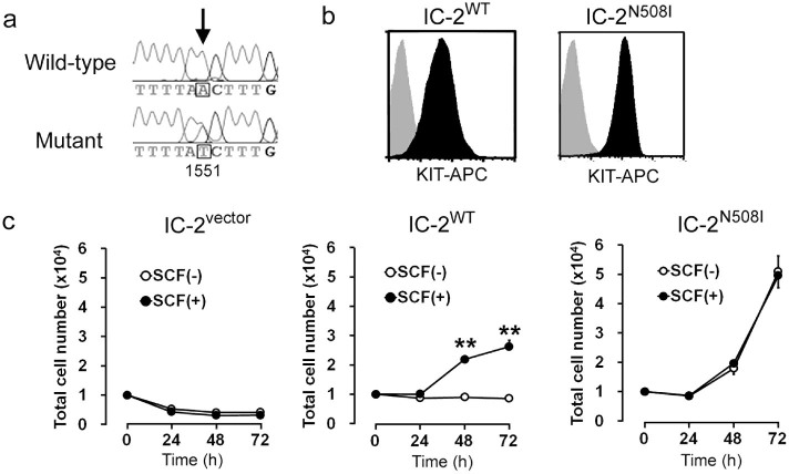
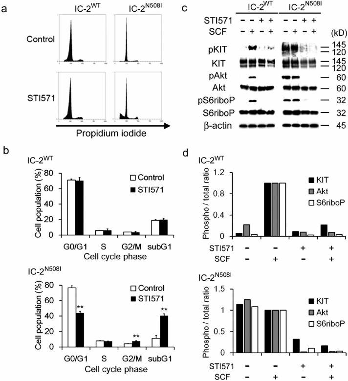
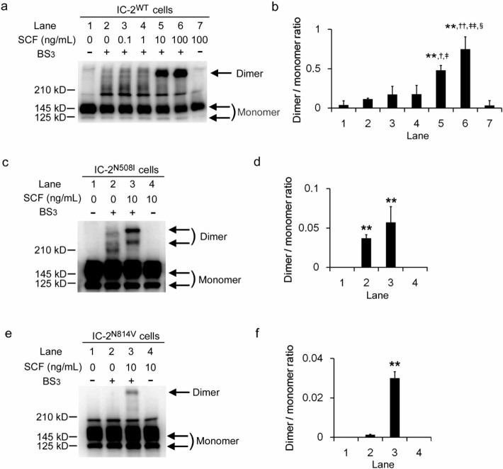
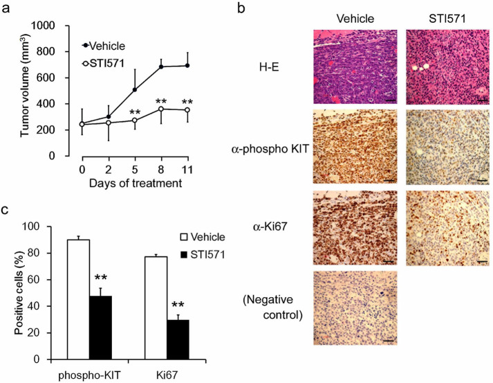
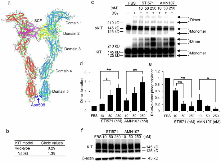
Mutations in the juxtamembrane and tyrosine kinase domains of the KIT receptor have been implicated in several cancers and are known to promote tumorigenesis. However, the pathophysiological manifestations of mutations in the extracellular domain remain unknown. In this study, we examined the impact of a mutation in the extracellular domain of KIT on mast cell tumorigenesis. A KIT mutant with an Asn508Ile variation (N508I) in the extracellular domain derived from a canine mast cell tumor was introduced into IC-2 cells. The IC-2(N508I) cells proliferated in a cytokine-independent manner and showed KIT auto-phosphorylation. Subcutaneous injection of IC-2(N508I) cells into the dorsal area of immunodeficient BALB/c-nu/nu mice resulted in the formation of solid tumors, but tumor progression was abrogated by treatment with a tyrosine kinase inhibitor (STI571). In addition, the N508I mutant KIT protein dimerized in the absence of the natural ligand, stem cell factor. Structure modeling indicates that the increased hydrophobicity of the mutant led to the stabilization of KIT dimers. These results suggest that this extracellular domain mutation confers a ligand-independent tumorigenic phenotype to mast cells by KIT auto-dimerization that is STI571-sensitive. This is the first report demonstrating the tumorigenic potential of a mutation in the extracellular domain of KIT.
Figures
Similar articles
-
Phosphatidylinositol 3'-kinase is required for growth of mast cells expressing the kit catalytic domain mutant.Cancer Res. 2003 Aug 1;63(15):4412-9. Cancer Res. 2003. PMID: 12907613
-
Transforming and differentiation-inducing potential of constitutively activated c-kit mutant genes in the IC-2 murine interleukin-3-dependent mast cell line.Am J Pathol. 1996 Jan;148(1):189-200. Am J Pathol. 1996. PMID: 8546206 Free PMC article.
-
Clustering of activating mutations in c-KIT's juxtamembrane coding region in canine mast cell neoplasms.J Invest Dermatol. 1999 Feb;112(2):165-70. doi: 10.1046/j.1523-1747.1999.00488.x. J Invest Dermatol. 1999. PMID: 9989791
-
Role of c-kit receptor tyrosine kinase in the development, survival and neoplastic transformation of mast cells.Pathol Int. 1996 Dec;46(12):933-8. doi: 10.1111/j.1440-1827.1996.tb03571.x. Pathol Int. 1996. PMID: 9110344 Review.
-
Role of c-kit/SCF in cause and treatment of gastrointestinal stromal tumors (GIST).Gene. 2007 Oct 15;401(1-2):38-45. doi: 10.1016/j.gene.2007.06.017. Epub 2007 Jul 3. Gene. 2007. PMID: 17659849 Review.
Cited by
-
Volleying plasma membrane proteins from birth to death: Role of J-domain proteins.Front Mol Biosci. 2022 Dec 15;9:1072242. doi: 10.3389/fmolb.2022.1072242. eCollection 2022. Front Mol Biosci. 2022. PMID: 36589230 Free PMC article. Review.
-
Effective targeting of PDGFRA-altered high-grade glioma with avapritinib.Cancer Cell. 2025 Apr 14;43(4):740-756.e8. doi: 10.1016/j.ccell.2025.02.018. Epub 2025 Mar 13. Cancer Cell. 2025. PMID: 40086436
-
Activating Mutation in the Receptor Tyrosine Kinase FLT3 with Clinicopathological Relevance in Canine Mast Cell Tumors.Vet Med Int. 2022 Aug 28;2022:9509900. doi: 10.1155/2022/9509900. eCollection 2022. Vet Med Int. 2022. PMID: 36072760 Free PMC article.
-
Mast Cells as Important Regulators in Autoimmunity and Cancer Development.Front Cell Dev Biol. 2021 Oct 12;9:752350. doi: 10.3389/fcell.2021.752350. eCollection 2021. Front Cell Dev Biol. 2021. PMID: 34712668 Free PMC article. Review.
-
A Canine c-kit Novel Mutation Isolated from a Gastrointestinal Stromal Tumor (GIST) Retains the Ability to Form Dimers but Lacks Autophosphorylation.Animals (Basel). 2025 May 16;15(10):1444. doi: 10.3390/ani15101444. Animals (Basel). 2025. PMID: 40427321 Free PMC article.
References
-
- Broxmeyer H. E. et al. The kit receptor and its ligand, steel factor, as regulators of hemopoiesis. Cancer Cells 3, 480–487 (1991). - PubMed
-
- Halaban R. Growth factors and tyrosine protein kinases in normal and malignant melanocytes. Cancer Metastasis Rev. 10, 129–140 (1991). - PubMed
-
- Huizinga J. D. et al. W/kit gene required for interstitial cells of Cajal and for intestinal pacemaker activity. Nature 373, 347–349 (1995). - PubMed
-
- Tsai M., Chen R., Tam S., Blenis J. & Galli S. J. Activation of MAP kinases, pp90rsk and pp70-S6 kinases in mouse mast cells by signaling through the c-kit receptor tyrosine kinase or FcRI: rapamycin inhibits activation of pp70-S6 kinase and proliferation in mouse mast cells. Eur. J. Immunol. 23, 3286–3291 (1993). - PubMed
Publication types
MeSH terms
Substances
LinkOut - more resources
Full Text Sources
Other Literature Sources
Research Materials
