

Age-related changes in monocytes exacerbate neointimal hyperplasia after vascular injury
- PMID: 25965835
- PMCID: PMC4627291
- DOI: 10.18632/oncotarget.3881
Age-related changes in monocytes exacerbate neointimal hyperplasia after vascular injury
Abstract
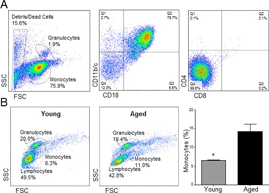
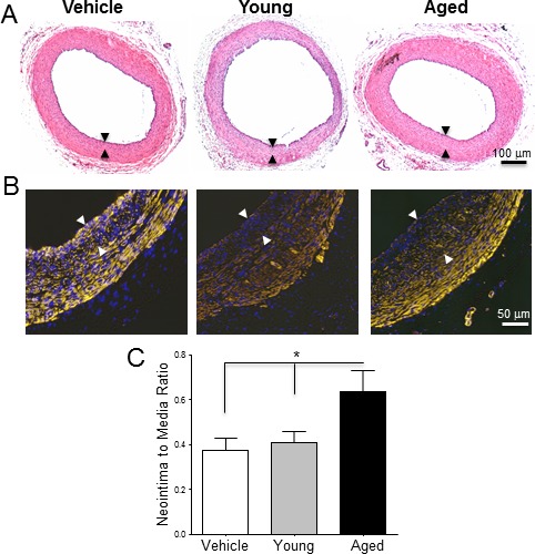
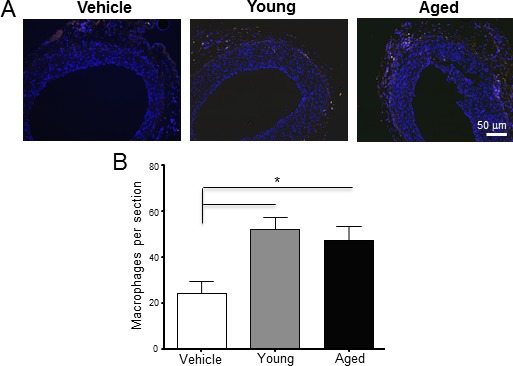
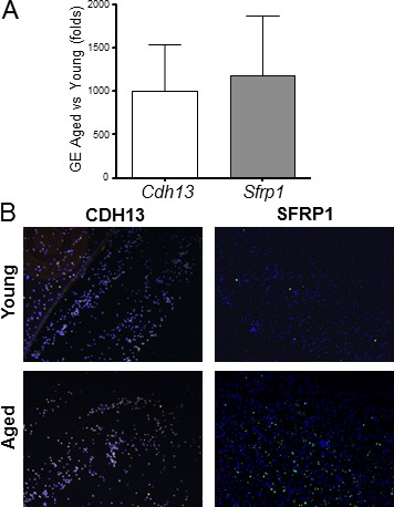
Neointimal hyperplasia is the leading cause of restenosis after endovascular interventions. It is characterized by the accumulation of myofibroblast-like cells and extracellular matrix in the innermost layer of the wall and is exacerbated by inflammation. Monocytes from either young or aged rats were applied perivascularly to injured vascular walls of young recipient animals. Monocytes from aged rats, but not young donors, increased neointima thickness. Accordingly, the gene expression profiles of CD11b+ monocytes from aged rats showed significant up-regulation of genes involved in cellular adhesion, lipid degradation, cytotoxicity, differentiation, and inflammation. These included cadherin 13 (Cdh13), colony stimulating factor 1 (Csf1), chemokine C-X-C motif ligand 1 (Cxcl1), endothelial cell-selective adhesion molecule (Esam), and interferon gamma (Ifng). In conclusion, our results suggest that the increased inflammatory and adhesive profile of monocytes contributes to pathological wall remodeling in aged-related vascular diseases.
Keywords: age; balloon injury; gene expression; monocytes; neointimal hyperplasia.
Conflict of interest statement
The authors indicate no potential conflicts of interests.
Figures
References
-
- Hokimoto S, Oike Y, Saito T, Kitaoka M, Oshima S, Noda K, Moriyama Y, Ishibashi F, Ogawa H. Increased expression of monocyte chemoattractant protein-1 in atherectomy specimens from patients with restenosis after percutaneous transluminal coronary angioplasty. Circ J. 2002;66:114–6. - PubMed
-
- Pietersma A, Kofflard M, de Wit LE, Stijnen T, Koster JF, Serruys PW, Sluiter W. Late lumen loss after coronary angioplasty is associated with the activation status of circulating phagocytes before treatment. Circulation. 1995;91:1320–5. - PubMed
-
- Fukuda D, Shimada K, Tanaka A, Kawarabayashi T, Yoshiyama M, Yoshikawa J. Circulating monocytes and in-stent neointima after coronary stent implantation. J Am Coll Cardiol. 2004;43:18–23. - PubMed
-
- Maddaluno M, Di Lauro M, Di Pascale A, Santamaria R, Guglielmotti A, Grassia G, Ialenti A. Monocyte chemotactic protein-3 induces human coronary smooth muscle cell proliferation. Atherosclerosis. 2011;217:113–9. - PubMed
-
- Kusaba K, Kai H, Koga M, Takayama N, Ikeda A, Yasukawa H, Seki Y, Egashira K, Imaizumi T. Inhibition of intrinsic interferon-gamma function prevents neointima formation after balloon injury. Hypertension. 2007;49:909–15. - PubMed
Publication types
MeSH terms
Grants and funding
LinkOut - more resources
Full Text Sources
Other Literature Sources
Medical
Research Materials
Miscellaneous
