

In vivo and in vitro evidence that intrinsic upper- and lower-limb skeletal muscle function is unaffected by ageing and disuse in oldest-old humans
- PMID: 25965867
- PMCID: PMC4516639
- DOI: 10.1111/apha.12524
In vivo and in vitro evidence that intrinsic upper- and lower-limb skeletal muscle function is unaffected by ageing and disuse in oldest-old humans
Abstract
Aim: To parse out the impact of advanced ageing and disuse on skeletal muscle function, we utilized both in vivo and in vitro techniques to comprehensively assess upper- and lower-limb muscle contractile properties in 8 young (YG; 25 ± 6 years) and 8 oldest-old mobile (OM; 87 ± 5 years) and 8 immobile (OI; 88 ± 4 years) women.
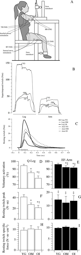
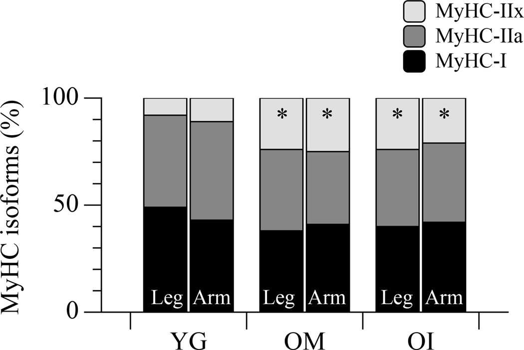
Methods: In vivo, maximal voluntary contraction (MVC), electrically evoked resting twitch force (RT), and physiological cross-sectional area (PCSA) of the quadriceps and elbow flexors were assessed. Muscle biopsies of the vastus lateralis and biceps brachii facilitated the in vitro assessment of single fibre-specific tension (Po).
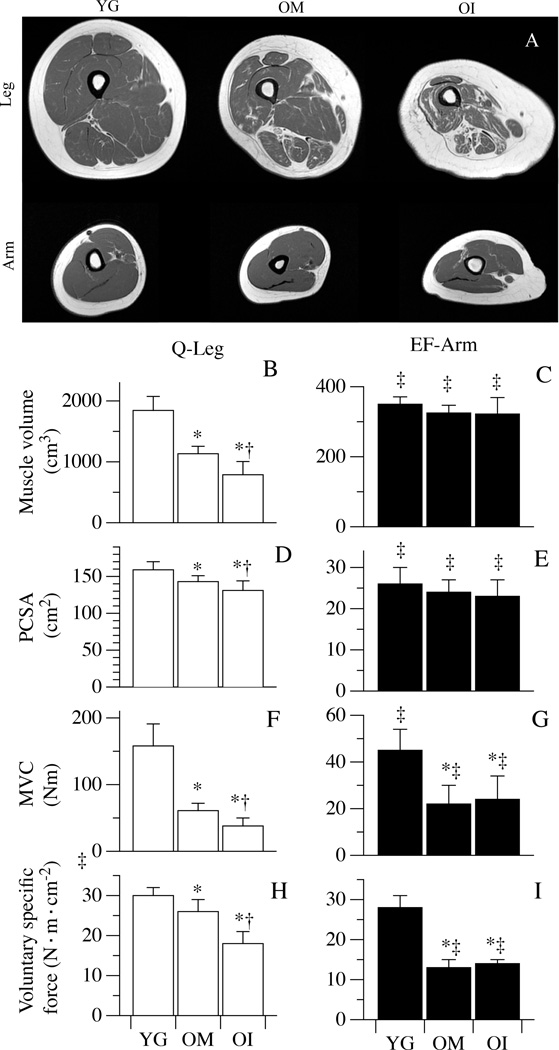
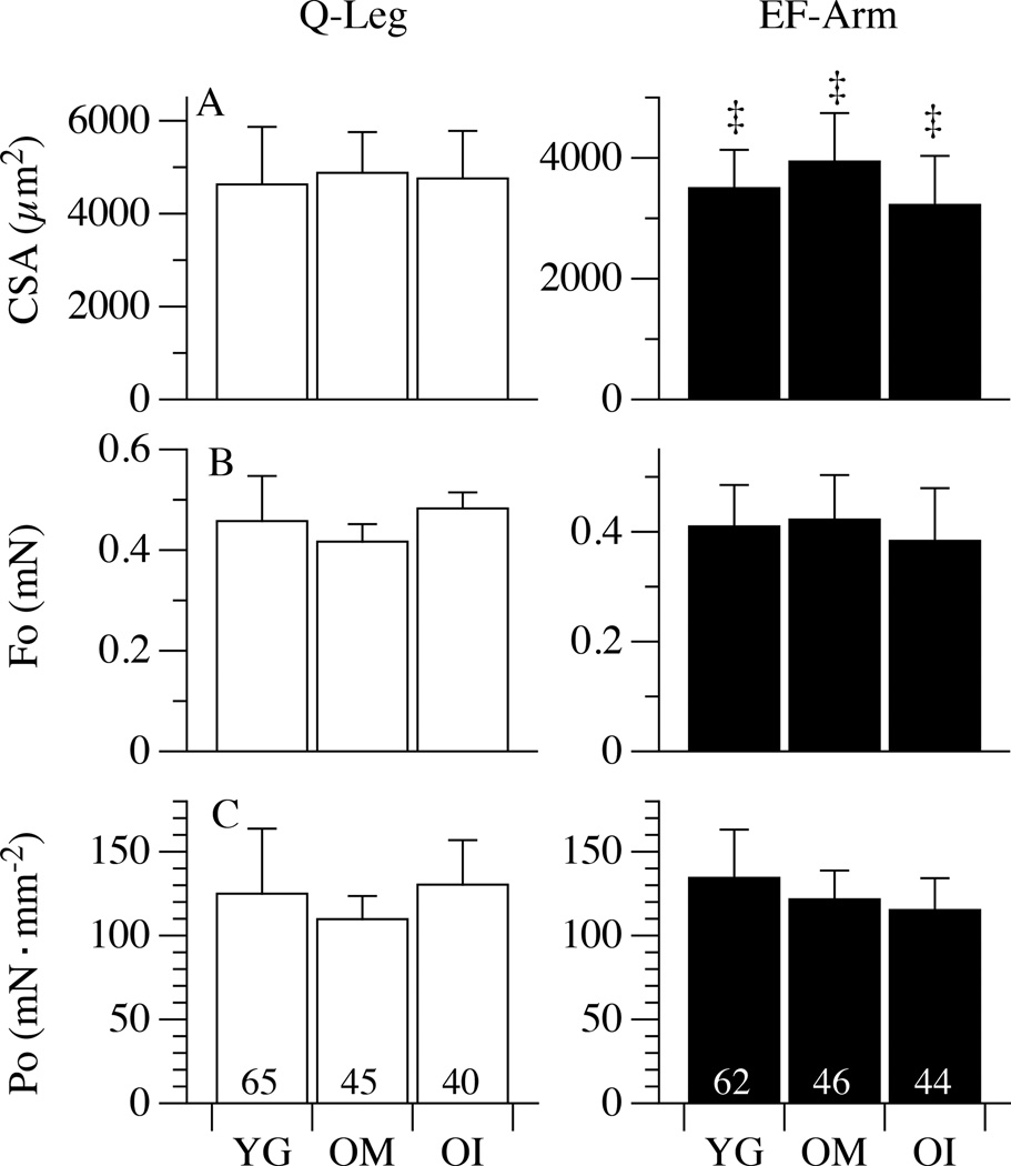
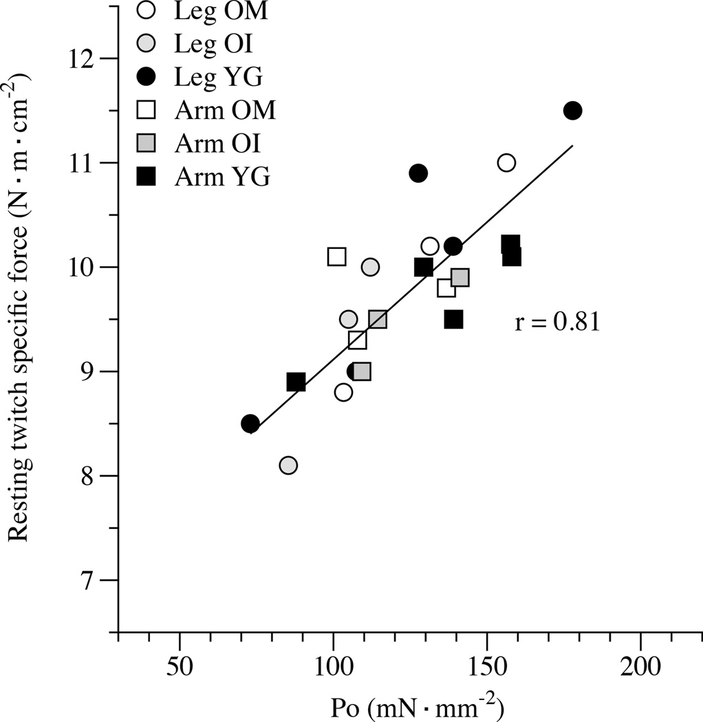
Results: In vivo, compared to the young, both the OM and OI exhibited a more pronounced loss of MVC in the lower limb [OM (-60%) and OI (-75%)] than the upper limb (OM = -51%; OI = -47%). Taking into account the reduction in muscle PCSA (OM = -10%; OI = -18%), only evident in the lower limb, by calculating voluntary muscle-specific force, the lower limb of the OI (-40%) was more compromised than the OM (-13%). However, in vivo, RT in both upper and lower limbs (approx. 9.8 N m cm(-2) ) and Po (approx. 123 mN mm(-2) ), assessed in vitro, implies preserved intrinsic contractile function in all muscles of the oldest-old and were well correlated (r = 0.81).
Conclusion: These findings suggest that in the oldest-old, neither advanced ageing nor disuse, per se, impacts intrinsic skeletal muscle function, as assessed in vitro. However, in vivo, muscle function is attenuated by age and exacerbated by disuse, implicating factors other than skeletal muscle, such as neuromuscular control, in this diminution of function.
Keywords: in vitro; in vivo; oldest-old; sarcopenia.
© 2015 Scandinavian Physiological Society. Published by John Wiley & Sons Ltd.
Conflict of interest statement
There is no conflict of interest.
Figures
Comment in
-
What is the mechanism for in vivo loss of skeletal muscle function in elderly women?Acta Physiol (Oxf). 2015 Sep;215(1):9-12. doi: 10.1111/apha.12547. Epub 2015 Jul 24. Acta Physiol (Oxf). 2015. PMID: 26132503 No abstract available.
References
-
- Aagaard P, Suetta C, Caserotti P, Magnusson SP, Kjaer M. Role of the nervous system in sarcopenia and muscle atrophy with aging: strength training as a countermeasure. Scand J Med Sci Sports. 2010;20:49–64. - PubMed
-
- Barns M, Gondro C, Tellam RL, Radley-Crabb HG, Grounds MD, Shavlakadze T. Molecular analyses provide insight into mechanisms underlying sarcopenia and myofibre denervation in old skeletal muscles of mice. Int J Biochem Cell Biol. 2014;53:174–185. - PubMed
-
- Berg HE, Larsson L, Tesch PA. Lower limb skeletal muscle function after 6 wk of bed rest. J Appl Physiol. 1997;82:182–188. - PubMed
-
- Blaauw B, Schiaffino S, Reggiani C. Mechanisms modulating skeletal muscle phenotype. Compr Physiol. 2013;3:1645–1687. - PubMed
-
- Bottinelli R, Pellegrino MA, Canepari M, Rossi R, Reggiani C. Specific contributions of various muscle fibre types to human muscle performance: an in vitro study. J Electromyogr Kinesiol. 1999;9:87–95. - PubMed
Publication types
MeSH terms
Grants and funding
LinkOut - more resources
Full Text Sources
Other Literature Sources
