. 2015 May 12;10(5):e0125219.
doi: 10.1371/journal.pone.0125219. eCollection 2015.

BMP-2 Is Involved in Scleral Remodeling in Myopia Development

Affiliations

BMP-2 Is Involved in Scleral Remodeling in Myopia Development

Honghui Li et al. PLoS One. .

Abstract

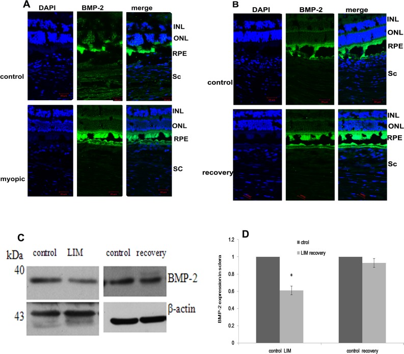
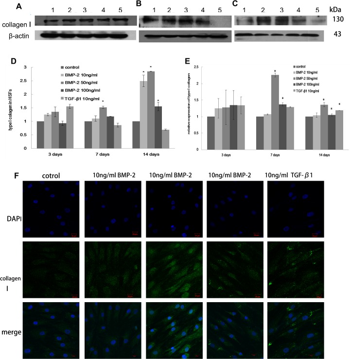
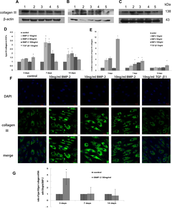
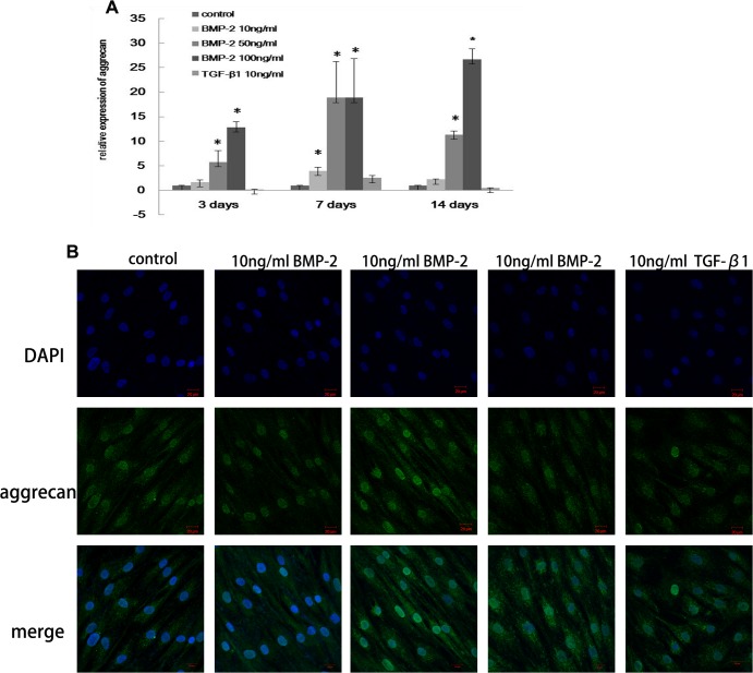
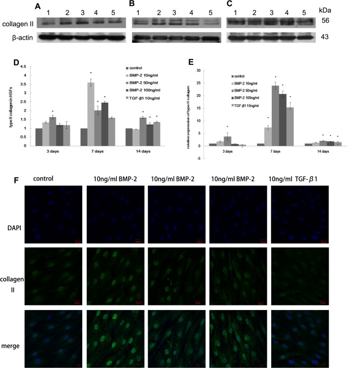
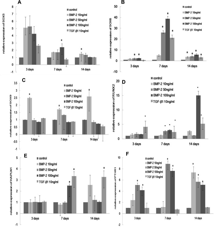
The development of myopia is associated with scleral remodeling, but it is unclear which factors regulate this process. This study investigated bone morphogenetic protein-2 (BMP-2) expression in the sclera of guinea pigs with lens-induced myopia (LIM) and after recovery from myopia and evaluated the effect of BMP-2 on extracellular matrix (ECM) synthesis in human scleral fibroblasts (HSFs) cultured in vitro. Lens-induced myopia was brought about in two groups of guinea pigs (the lens-induced myopia and myopia recovery groups) by placing -4.00 D lenses on the right eye for three weeks. The left eye served as a contralateral control. In the recovery group, the lenses were removed after one week. The refractive power and axial length of the eyes were measured, and the BMP-2 expression levels in the sclera were measured. After three weeks, the lens-induced eyes acquired relative myopia in both groups of guinea pigs. Immunostaining of the eyeballs revealed significantly decreased BMP-2 expression in the posterior sclera of the myopic eyes compared to the contralateral eyes. One week after lens removal, BMP-2 expression recovered, and no differences were observed between the experimental and contralateral eyes in the recovery group. HSFs were cultured with BMP-2 or transforming growth factor-β1 (TGF-β1). Type I and type III collagen synthesis was significantly up-regulated following BMP-2 treatment in culture after one and two weeks, but the ratio of type III to type I collagen mRNA was not increased. Biosynthesis of glycosaminoglycan (GAG) and aggrecan was increased in HSFs treated with BMP-2. Some chondrogenesis-associated genes expression increased in HSFs treated with BMP-2. From this study, we concluded that BMP-2 is involved in scleral remodeling in the development and recovery of lens-induced myopia.


Conflict of interest statement

Competing Interests: The authors have declared that no competing interests exist.

Figures

Fig 1
Fig 1. BMP-2 expression in myopia and recovery from myopia of guinea pigs.
Immunofluorescence analysis of BMP-2 expression in the retina, RPE, choroid and sclera of guinea pigs. The staining in the myopic sclera of the LIM group was weaker than that of the control eyes (A). Changes in the expression of BMP-2 in the sclera of guinea pigs detected by western blot analysis (C,D). The data are expressed as the mean±SEM, * p≤0.05 vs. the contralateral eye. INL inner nuclear layer, ONL outer nuclear layer, RPE retinal pigmental epithelium, chl choroid, Sc sclera layer.
Fig 2
Fig 2. The effect of BMP-2 on type I collagen expression in HSFs.
The synthesis of type I collagen in HSFs after 3 days (A), 1 week (B) and 2 weeks (C) of culture. Lane 1-Untreated control, Lane 2–10 ng/ml BMP-2, Lane 3- 50ng/ml BMP-2, Lane 4–100 ng/ml BMP-2, Lane 5- 10ng/ml TGF-β1. The data are expressed as the mean±SEM, N = 3, * p≤0.05 vs. control (D). The mRNA level of type I collagen in HSFs is shown (E). The data are expressed as the mean±SEM, N = 3, * p≤0.05 vs. control. Immunocytochemistry (F) also shows a BMP-2 induced increase in collagen type I expression in HSFs after 7 days culture.
Fig 3
Fig 3. The effect of BMP-2 on type I collagen expression in HSFs.
The expression of type III collagen in HSFs after 3 days (A), 1 week (B) and 2 weeks (C). Lane 1-Untreated control, Lane 2–10 ng/ml BMP-2, Lane 3–50 ng/ml BMP-2, Lane 4–100 ng/ml BMP-2, Lane 5–10 ng/ml TGF-β1. The data are expressed as the mean±SEM, N = 3, * p≤0.05 vs. control. Real-time PCR (E) also shows an increase in type III collagen after BMP-2 and TGF-β1 treatment (D). Immunocytochemistry (F) reveals the expression of type III collagen in HSFs after 7 days culture. The ratio of type III/I collagen mRNA increased significantly in HSFs treated with 50ng/BMP-2 for 3 days compared to control cells, but this ratio decreased and was not changed after 1 or 2 weeks of treatment (G). The data are expressed as the mean±SEM. N = 3, * p≤0.05 vs. control.
Fig 4
Fig 4. The effect of BMP-2 on GAG synthesis in HSFs.
GAG synthesis was visualized by toluidine blue staining of HSFs in culture. GAG was observed after treatment with BMP-2 for one week but not in control cells. Original magnification: 40×.
Fig 5
Fig 5. The effect of BMP-2 on aggrecan expression in HSFs.
Aggrecan mRNA level in HSFs was analyzed after 3 days, 1 week and 2 weeks (A). Immunocytochemistry shows an increase in aggrecan expression after 7 days (B). The data are expressed as the mean±SEM. N = 3, * p≤0.05 vs. Control.
Fig 6
Fig 6. The effect of BMP-2 on type II collagen expression in HSFs.
Type II collagen synthesis in HSFs after 3 days (A), 1 week (B), 2 weeks (C) detected by western lot analysis (D). Lane 1- Untreated control, Lane 2–10 ng/ml BMP-2, Lane 3–50 ng/ml BMP-2, Lane 4–100 ng/ml BMP-2, Lane 5–10 ng/ml TGF-β1. The expression of type II collagen mRNA was analyzed using real-time PCR (E). Immunocytochemistry showed an increase in collagen II expression in HSFs after 7 days of treatment with BMP-2 and TGF-β1 in culture (F). The data are expressed as the mean±SEM. N = 3, * p≤0.05 vs. Control.
Fig 7
Fig 7. The effect of BMP-2 on chondrogenesis-related genes expression in HSFs.
The mRNA level of SOX5 (A), SOX6 (B), SOX9 (C), RUNX2 (D), HAPLN (E), PTHR1 (F) was analyzed. HSFs were treated for 3 days, 1 week, and 2 weeks seperately. *: Denotes a significant difference relative to the control group, P< 0.05.

Similar articles

Cited by

References

    1. Lin LL, Shih YF, Tsai CB, Chen CJ, Lee LA, Hung PT, et al. Epidemiologic study of ocular refraction among schoolchildren in Taiwan in 1995. 1999; Optom Vis Sci 76: 275–281. - PubMed
    1. Goh WS, Lam CS. Changes in refractive trends and optical components of Hong Kong Chinese aged 19–39 years. 1994; Ophthalmic Physiol Opt 14: 378–382. - PubMed
    1. Grey RH, Burns-Cox CJ, Hughes A. Blind and partial sight registration in Avon. 1989; Br J Ophthalmol 73: 88–94. - PMC - PubMed
    1. Sperduto RD, Seigel D, Roberts J, Rowland M. Prevalence of myopia in the United States. 1983; Arch Ophthalmol 101: 405–407. - PubMed
    1. Leng Y, Lan W, Yu K, Liu B, Yang Z, Li Z, et al. Effects of confined space and near vision stimulation on refractive status and vitreous chamber depth in adolescent rhesus monkeys. 2010; Sci China Life Sci 53: 1433–1439. 10.1007/s11427-010-4099-9 - DOI - PubMed

Publication types

Substances