Review
. 2015 Mar;35(2):145-55.
doi: 10.1016/j.semnephrol.2015.02.004.

Endothelin and the renal microcirculation

Affiliations
Review

Endothelin and the renal microcirculation

Zhengrong Guan et al. Semin Nephrol. 2015 Mar.

Abstract

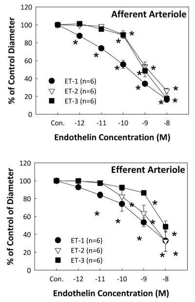
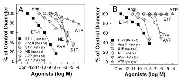
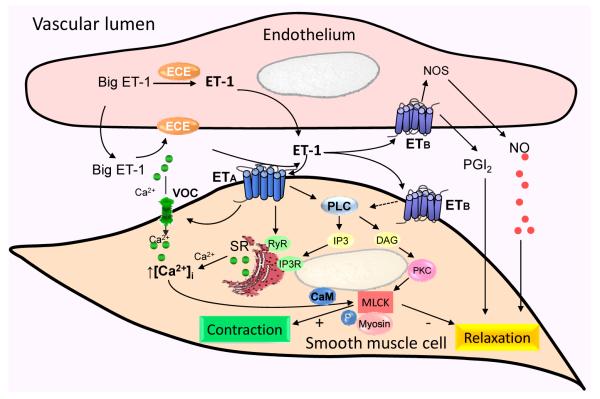
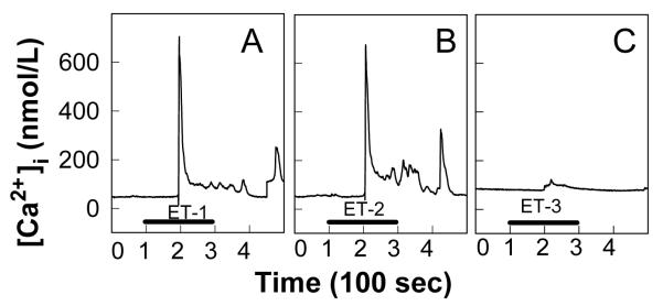
Endothelin (ET) is one of the most potent renal vasoconstrictors. Endothelin plays an essential role in the regulation of renal blood flow, glomerular filtration, sodium and water transport, and acid-base balance. ET-1, ET-2, and ET-3 are the three distinct endothelin isoforms comprising the endothelin family. ET-1 is the major physiologically relevant peptide and exerts its biological activity through two G-protein-coupled receptors: ET(A) and ET(B). Both ET(A) and ET(B) are expressed by the renal vasculature. Although ET(A) are expressed mainly by vascular smooth muscle cells, ET(B) are expressed by both renal endothelial and vascular smooth muscle cells. Activation of the endothelin system, or overexpression of downstream endothelin signaling pathways, has been implicated in several pathophysiological conditions including hypertension, acute kidney injury, diabetic nephropathy, and immune nephritis. In this review, we focus on the effects of endothelin on the renal microvasculature, and update recent findings on endothelin in the regulation of renal hemodynamics.

Keywords: Kidney; afferent arteriole; autoregulation; efferent arteriole; glomerular filtration.


Figures

Figure 1
Figure 1
Renal microvascular responses to endothelins Kidneys were superfused with increasing concentrations of ET-1, ET-2 or ET-3 from 10−12 to 10−8 M (5 minutes at each concentration) using the in vitro blood-perfused juxtamedullary nephron preparation while perfusion pressure was maintained at 100 mmHg. Afferent arteriolar responses (A) and efferent arteriolar responses (B) were measured at 12-second intervals and calculated from the average of all measurements obtained during the final 2 minutes of each 5-minute period. Data are expressed as the percent of control diameter. Values are mean ± SEM. *P<0.05 vs. control diameter in same group. Figure is modified from.
Figure 2
Figure 2
Comparison of effect of ET-1 with other common vasoconstrictors on afferent and efferent arteriolar reactivity in the rat juxtamedullary nephron preparation A: the vasoconstriction of afferent arterioles to ET-1 (square symbols) is greater than the vasoconstriction induced by other GPCR agonists, angiotensin II (Ang II),, arginine vasopressin (AVP), norepinephrine (NE), sphingosine-1-phosphate (S1P), and ATP. B: the vasoconstriction of efferent arterioles to ET-1 (square symbols) is also greater than the vasoconstriction evoked by Ang II, AVP, and NE while efferent arterioles did not show any detectable responses to S1P and ATP. All of the studies were conducted using the rat juxtamedullary nephron preparation.
Figure 3
Figure 3
Possible intracellular signaling pathways for ET in the renal microvasculature ET-1 is produced from big ET-1 catabolized by the ET-converting enzyme (ECE) expressed in endothelial cells. The majority of ET-1 is released towards the basolateral side of the endothelium. ET-1 acts in a paracrine/autocrine manner to influence endothelial or vascular smooth muscle cell function via activation of ETA and ETB receptors. Activation of ETA receptors in vascular smooth muscle cells increases cytosolic calcium concentration ([Ca2+]i) by activating voltage-gated calcium channels (VOC), mobilization of Ca2+ from the sarcoplasmic reticulum (SR) by activating phospholipase-C (PLC), diacylglycerol (DAG) and inositol triphosphate (IP3) as well as cyclic adenine diphosphate ribose (cADPR) cyclase/ ryanodine receptor (RyR) pathways. Activation of endothelial ETB receptors releases nitric oxide (NO) and prostaglandins (PGs) leading to vasodilation. Activation of ETB receptors in vascular smooth muscle cells mediates vasoconstriction through undefined intracellular mechanisms (dashed arrow). PKC, protein kinase-C; CaM, calmodulin; MLCK, myosin light chain kinase; MLCK; +, stimulate; -, inhibit.
Figure 4
Figure 4
Effect of ETs on cytosolic Ca2+ concentration in single rat preglomerular microvascular smooth muscle cells Typical cytosolic Ca2+ concentration ([Ca2+]i) recordings obtained from single smooth muscle cells isolated from rat preglomerular microvessels in response to 100 nmol/L ET-1, ET-2, and ET-3 are depicted in panels A, B, and C, respectively. The black bar indicates the period of ET peptide administration. The time between two major ticks in X axis represents 100 second intervals. Figure is modified from.

References

    1. Hickey KA, Rubanyi G, Paul RJ, Highsmith RF. Characterization of a coronary vasoconstrictor produced by cultured endothelial cells. Am J Physiol. 1985;248(5 Pt 1):C550–6. - PubMed
    1. Yanagisawa M, Kurihara H, Kimura S, Tomobe Y, Kobayashi M, Mitsui Y, Yazaki Y, Goto K, Masaki T. A novel potent vasoconstrictor peptide produced by vascular endothelial cells. Nature. 1988;332(6163):411–5. - PubMed
    1. Itoh Y, Yanagisawa M, Ohkubo S, Kimura C, Kosaka T, Inoue A, Ishida N, Mitsui Y, Onda H, Fujino M, et al. Cloning and sequence analysis of cDNA encoding the precursor of a human endothelium-derived vasoconstrictor peptide, endothelin: identity of human and porcine endothelin. FEBS Lett. 1988;231(2):440–4. - PubMed
    1. Russell FD, Davenport AP. Secretory pathways in endothelin synthesis. Br J Pharmacol. 1999;126(2):391–8. - PMC - PubMed
    1. Johnstrom P, Fryer TD, Richards HK, Maguire JJ, Clark JC, Pickard JD, Davenport AP. Positron emission tomography of [18F]-big endothelin-1 reveals renal excretion but tissue-specific conversion to [18F]-endothelin-1 in lung and liver. Br J Pharmacol. 2010;159(4):812–9. - PMC - PubMed

Publication types

MeSH terms

LinkOut - more resources