. 2015 May 12:5:10038.
doi: 10.1038/srep10038.

A method for human teratogen detection by geometrically confined cell differentiation and migration

Affiliations

A method for human teratogen detection by geometrically confined cell differentiation and migration

Jiangwa Xing et al. Sci Rep. .

Erratum in

Abstract

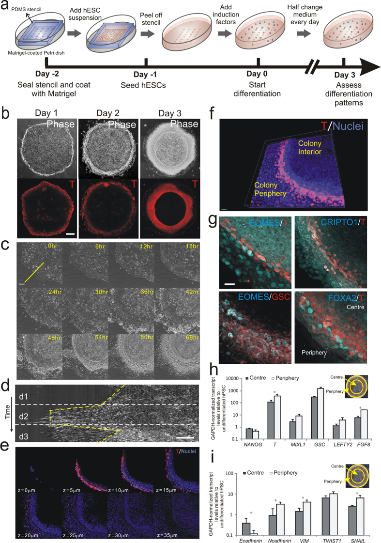
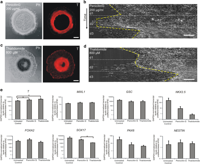
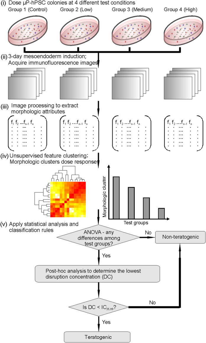
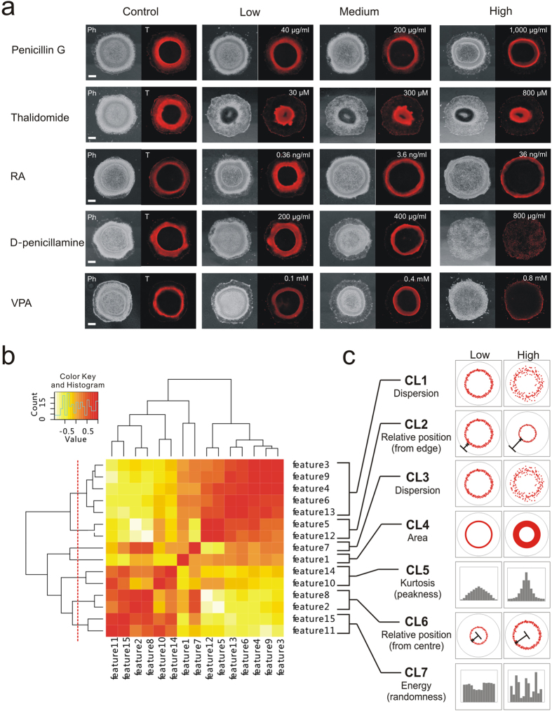
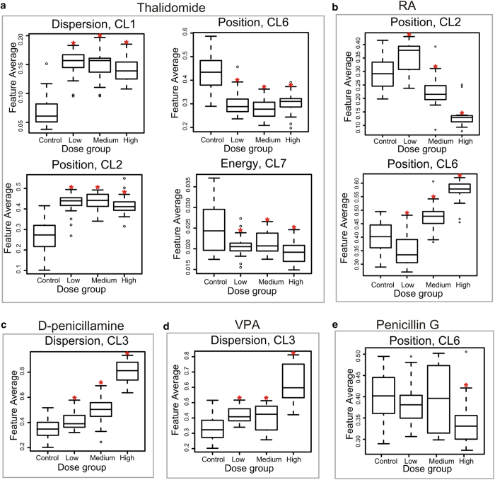
Unintended exposure to teratogenic compounds can lead to various birth defects; however current animal-based testing is limited by time, cost and high inter-species variability. Here, we developed a human-relevant in vitro model, which recapitulated two cellular events characteristic of embryogenesis, to identify potentially teratogenic compounds. We spatially directed mesoendoderm differentiation, epithelial-mesenchymal transition and the ensuing cell migration in micropatterned human pluripotent stem cell (hPSC) colonies to collectively form an annular mesoendoderm pattern. Teratogens could disrupt the two cellular processes to alter the morphology of the mesoendoderm pattern. Image processing and statistical algorithms were developed to quantify and classify the compounds' teratogenic potential. We not only could measure dose-dependent effects but also correctly classify species-specific drug (Thalidomide) and false negative drug (D-penicillamine) in the conventional mouse embryonic stem cell test. This model offers a scalable screening platform to mitigate the risks of teratogen exposures in human.


Figures

Figure 1
Figure 1. Formation of annular mesoendoderm pattern in μP-hPSC colony.
(a) Schematic depicting micropatterning of hPSC colonies and mesoendoderm induction. (b) Phase and immunofluorescence images of mesoendoderm marker Brachyury (T) 1–3 days post mesoendoderm induction. Scale bar, 200 μm. (c) Montage from a 3-day phase imaging on a quarter section of a circular μP-hPSC colony. Scale bar, 100 μm. (d) Kymograph analysis showing the movement of cells along the yellow line shown in (c) throughout the 3-day live imaging time frame. Scale bar, 50 μm. (e, f) Confocal z-stack sections of T (red) and cell nuclei (blue)-labeled multicellular annular structure (e) and its 3-D reconstruction image (f) on day 3. Scale bar, 30 μm in (f). (g) Immunofluorescence images of mesoendoderm markers EOMES, CRIPTO1 GSC and FOXA2. Scale bar, 20 μm. (h,i) Gene expression levels of mesoendoderm markers (h) and EMT markers (g) in colony centre and periphery on day 3 relative to undifferentiated hPSCs. Data are average ± s.d. of three experiments with duplicate samples. *, p < 0.05 in paired t-test. Insets, phase image showing colony periphery and centre.
Figure 2
Figure 2. Disruption of annular mesoendoderm pattern by teratogen treatment.
(a,c) Phase and T fluorescence images of μP-hPSC colonies under Penicillin G (a) and Thalidomide (c) treatment after 3-day mesoendoderm induction. Scale bar, 200 μm. (b,d) Kymographs of cell movements around colony periphery during 3-day mesoendoderm induction under Penicillin G (b) and Thalidomide (d) treatment. Scale bar, 50 μm. (e) RT-PCR analysis of expression levels of germ layer markers in untreated, Penicillin G-treated and Thalidomide-treated colonies on day 3 relative to undifferentiated hPSCs. Mesoendoderm markers are T, MIXL1 and GSC; mesoderm marker is NKX2.5; definitive endoderm markers are FOXA2 and SOX17; and ectoderm markers are PAX6 and NESTIN. Data are average ± s.d. of three experiments with duplicate samples. *, p < 0.05 in paired t-test.
Figure 3
Figure 3. A quantitative morphometric assay for teratogen screening.
Workflow for using the μP-hPSC model to determine the teratogenic potential of a test compound. Disruption concentration (DC) refers to the lowest concentration which morphologically disrupts the mesoendoderm pattern.
Figure 4
Figure 4. Generation of morphologic clusters by unsupervised feature clustering.
(a) Phase and T immunofluorescence images of μP-hPSC colonies in different drug test groups on day 3. Scale bar, 200 μm. (b) Hierarchical clustering of morphologic attributes based on feature correlations. Dash line indicates that 7 clusters were acquired. (c) Graphical interpretations of the 7 morphologic clusters to describe changes to the annular mesoendoderm pattern.
Figure 5
Figure 5. Teratogen screening results in the μP-hPSC model.
(a-e) Boxplots of morphologic cluster readout, which showed clear dose-dependent disruption effects of each drug among the four test groups. The low, medium, high tested concentrations of each drug were 30 μM , 300 μM, 800 μM for Thalidomide in (a); 0.00036 μg/ml, 0.0036 μg/ml and 0.036 μg/ml for RA in (b); 200 μg/ml, 400 μg/ml and 800 μg/ml for D-penicillamine in (c); 0.1 mM, 0.4 mM, 0.8 mM for VPA in (d); 40 μg/ml, 200 μg/ml, and 1,000 μg/ml for Penicillin G in (e). *: p < 0.0083 in post-hoc analysis.

References

    1. Haschek W. M., Rousseaux C. G. & Wallig M. A. [Chapter 20 Developmental Pathology] Fundamentals of Toxicologic Pathology [634] Academic Press London, UK 2010).
    1. Wobus A. M. & Löser P. Present state and future perspectives of using pluripotent stem cells in toxicology research. Arch. Toxicol. 85, 79–117 (2011). - PMC - PubMed
    1. Bremer S., Pellizzer C., Hoffmann S., Seidle T. & Hartung T. The development of new concepts for assessing reproductive toxicity applicable to large scale toxicological programmes. Curr. Pharm. Des. 13, 3047–58 (2007). - PubMed
    1. Kameoka S., Babiarz J., Kolaja K. & Chiao E. A high-throughput screen for teratogens using human pluripotent stem cells. Toxicol. Sci. 137, 76–90 (2014). - PubMed
    1. Colleoni S. et al. Characterisation of a neural teratogenicity assay based on human ESCs differentiation following exposure to valproic acid. Curr. Med. Chem. 19, 6065–71 (2012). - PubMed

Publication types

MeSH terms

LinkOut - more resources