Review
. 2015;8(1):12-21.
doi: 10.2174/1874467208666150507103417.

Regulation of Cardiac Calcium Channels in the Fight-or-Flight Response

Affiliations
Review

Regulation of Cardiac Calcium Channels in the Fight-or-Flight Response

William A Catterall. Curr Mol Pharmacol. 2015.

Abstract

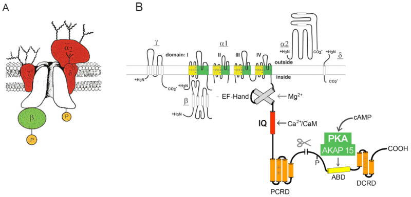
Intracellular calcium transients generated by activation of voltage-gated calcium (CaV) channels generate local signals, which initiate physiological processes such as secretion, synaptic transmission, and excitation-contraction coupling. Regulation of calcium entry through CaV channels is crucial for control of these physiological processes. In this article, I review experimental results that have emerged over several years showing that cardiac CaV1.2 channels form a local signaling complex, in which their proteolytically processed distal C-terminal domain, an A-Kinase Anchoring Protein, and cyclic AMP-dependent protein kinase (PKA) interact directly with the transmembrane core of the ion channel through the proximal C-terminal domain. This signaling complex is the substrate for β-adrenergic up-regulation of the CaV1.2 channel in the heart during the fight-or-flight response. Protein phosphorylation of two sites at the interface between the distal and proximal C-terminal domains contributes importantly to control of basal CaV1.2 channel activity, and phosphorylation of Ser1700 by PKA at that interface up-regulates CaV1.2 activity in response to β-adrenergic signaling. Thus, the intracellular C-terminal domain of CaV1.2 channels serves as a signaling platform, mediating beat-to-beat physiological regulation of channel activity and up-regulation by β-adrenergic signaling in the fight-or-flight response.


Conflict of interest statement

CONFLICT OF INTEREST

The author confirms that this article content has no conflict of interest.

Figures

Fig. 1
Fig. 1. The CaV1 calcium channel signaling complex
A. Subunit structure of the CaV1.1 channel from skeletal muscle transverse tubules. The pore-forming α1 subunit is illustrated in the center of the complex with interacting β, α2δ, and γ subunits. Modified from [27, 40, 80]. B. A transmembrane folding diagram illustrating the subunits of CaV1.2 channels. The C-terminal domain of the cardiac calcium CaV1.2 channels is shown in expanded presentation to illustrate the regulatory interactions clearly. ABD, AKAP15 binding domain; DCRD, distal C-terminal regulatory domain; PCRD, proximal C-terminal regulatory domain; scissors, site of proteolytic processing. An EF-hand motif involved in regulation of Mg2+ and an IQ-like motif involved in calcium-dependent inactivation are also illustrated.
Fig. 2
Fig. 2. Disruption of the AKAP15-leucine zipper interaction inhibits β-adrenergic receptor regulation of CaV1.2 channels in rat ventricular myocytes
(A), Left panel, representative currents elicited by 300-ms test pulses to 0 mV before (closed symbols) and after (open symbols) 5 min exposure to Iso (1 μM). Right panel, mean (± sem) current-voltage relationships before (closed symbols) and after (open symbols) 5 min exposure to isoproterenol in the absence of peptide dialysis. (B), the effect of intracellular dialysis with AKAP15LZ (38–54) (100 μM; n = 14, black triangles) on the response of ICa to isoproterenol as compared to control (circles and gray). Modified from [61].
Fig. 3
Fig. 3. Interaction of the cleaved distal C-terminus with truncated CaV1.2 channels and autoinhibition of channel activity
(A) Representative Ba2+ currents elicited by 20-ms test pulses from −80 to +20 mV recorded through full-length CaV1.2 (closed squares), CaV1.2 Δ1821 (closed circles), and CaV1.2Δ1821 plus distal1822-2171 (open circles) channels. (B) Mean (± sem) current-voltage relationships of full-length CaV1.2 (closed squares), CaV1.2Δ1821 (closed circles), and CaV1.2Δ1821 plus distal1822-2171 (open circles) channels. Peak currents at +10 mV were: −3.73 ± 0.29 nA/pC (n=10) for full-length; −10.75 ± 1.07 nA/pC for CaV1.2Δ1821, n = 11, p<0.001; and −0.55 ± 0.08 nA/pC, n=24 for CaV1.2Δ1821 plus distal1822-2171. Modified from [67].
Fig. 4
Fig. 4. Regulation of CaV1.2 channel activity by optimal expression of cDNA encoding CaV1.2 channel subunits and AKAP15
(A) Representative Ba2+ currents (Top) and current-voltage relationships (Bottom) of CaV1.2 channels expressed in tsA-201 cells by co-transfection of the indicated cDNAs encoding CaV1.2Δ1800, distal C-terminus (DCT), AKAP15, and 5 μM Forskolin (FSK). (B) Representative Ba2+ currents (Top) and current-voltage relationships (Bottom) of CaV1.2 channels expressed in tsA-201 cells by co-transfection of the indicated cDNAs encoding CaV1.2Δ1800, distal C-terminus (DCT), AKAP15, and 5 μM Forskolin (FSK) with wild-type CaV1.2Δ1800 and mutants S1700A, T1704A, and S1700A/T1704A. Mean±SEM; significance determined by ANOVA.
Fig. 5
Fig. 5. Reduced basal Ca2+ current and impaired response to β-adrenergic activation in adult cardiomyocytes
(A) ICa was recorded under basal conditions and 5 min after application of 10 nM Iso in WT and STAA cardiomyocytes. Basal current was stable for two min before addition of Iso. (B) Baseline subtracted Iso-induced increment in ICa density plotted against Iso concentration. *** p<0.001, WT vs. SA. *, p<0.05 SA vs STAA. Modified from [76, 77].
Fig. 6
Fig. 6. The docking model of the proximal and distal C-terminal domains in the CaV1.2 channel signaling complex
(A) Shown in ribbon representation with their α-helical regions colored. Side chains of R1696 and R1697 in the PCRD are shown in stick representation with nitrogen atoms in blue. Side chains of E2103, E2104, and E2106 in the DCRD are shown in stick representation with oxygen atoms in red. (B) Rotated view of the model in panel A showing the side chain of S1700 in stick representation with the oxygen atom in red.

References

    1. Reuter H. Properties of two inward membrane currents in the heart. Annu Rev Physiol. 1979;41:413–424. - PubMed
    1. Tsien RW. Calcium channels in excitable cell membranes. Annu Rev Physiol. 1983;45:341–358. - PubMed
    1. Bers DM. Cardiac excitation-contraction coupling. Nature. 2002;415:198–205. - PubMed
    1. Reuter H. Calcium channel modulation by neurotransmitters, enzymes and drugs. Nature. 1983;301:569–574. - PubMed
    1. Catterall WA. Excitation-contraction coupling in vertebrate skeletal muscle: A tale of two calcium channels. Cell. 1991;64:871–874. - PubMed

Publication types

Substances