. 2015 May 14:8:50.
doi: 10.1186/s13045-015-0146-0.

Long non-coding RNA ANRIL is upregulated in hepatocellular carcinoma and regulates cell apoptosis by epigenetic silencing of KLF2

Affiliations

Long non-coding RNA ANRIL is upregulated in hepatocellular carcinoma and regulates cell apoptosis by epigenetic silencing of KLF2

Ming-de Huang et al. J Hematol Oncol. .

Erratum in

Abstract

Background: Hepatocellular carcinoma (HCC) is one of the leading causes of cancer-related death, especially in China. And the mechanism of its progression remains poorly understood. Growing evidence indicates that long non-coding RNAs (lncRNAs) are found to be dysregulated in many cancers, including HCC. ANRIL, a lncRNA co-clustered mainly with p14/ARF has been reported to be dysregulated in gastric cancer, esophageal squamous cell carcinoma, and lung cancer. However, its clinical significance and potential role in HCC are still not documented.

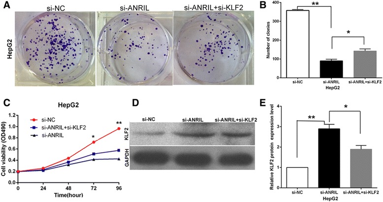
Methods and results: In this study, expression of ANRIL was analyzed in 77 HCC tissues and matched normal tissues by using quantitative polymerase chain reaction (qRT-PCR). ANRIL expression was upregulated in HCC tissues, and the higher expression of ANRIL was significantly correlated with tumor size and Barcelona Clinic Liver Cancer (BCLC) stage. Moreover, taking advantage of loss-of-function experiments in HCC cells, we found that knockdown of ANRIL expression could impair cell proliferation and invasion and induce cell apoptosis both in vitro and in vivo. We also found that ANRIL could epigenetically repress Kruppel-like factor 2 (KLF2) transcription in HCC cells by binding with PRC2 and recruiting it to the KLF2 promoter region. We also found that SP1 could regulate the expression of ANRIL.

Conclusion: Our results suggest that lncRNA ANRIL, as a growth regulator, may serve as a new biomarker and target for therapy in HCC.


Figures

Figure 1
Figure 1
Relative ANRIL expression in HCC tissues and HCC cell lines, and ANRIL regulation by SP1. (A) Relative ANRIL expression in HCC tissues (n = 77) compared with corresponding non-tumor tissues (n = 77). ANRIL expression was examined by qPCR and normalized to GAPDH expression. Results were presented as ΔCT in tumor tissues relative to normal tissues. (B) ANRIL expression was classified into two groups. Positive ΔΔCT meant high ANRIL expression. Negative ΔΔCT meant low ANRIL expression. (C) Relative ANRIL expression levels of HCC cell lines (HepG2, Hep3B, MHHC-97H) compared with those in the normal hepatic epithelium cell line (L02). (D) ChIP-qPCR of SP1 occupancy and binding in the ANRIL promoter in HepG2 and Hep3B cells, and IgG as a negative control. (E) The SP1 expression level was determined by qPCR when HepG2 cells were transfected with si-SP1. (F) The ANRIL expression level was determined by qPCR when HepG2 cells were transfected with si-SP1. (G) The SP1 expression level was determined by qPCR when Hep3B cells were transfected with si-SP1. (H) The ANRIL expression level was determined by qPCR when Hep3B cells were transfected with si-SP1. (I) The SP1 expression level was determined by qPCR when HepG2 cells were transfected with EGFP-SP1. (J) The ANRIL expression level was determined by qPCR when HepG2 cells were transfected with EGFP-SP1. (K) The SP1 expression level was determined by qPCR when Hep3B cells were transfected with EGFP-SP1. (L) The ANRIL expression level was determined by qPCR when Hep3B cells were transfected with EGFP-SP1. *P < 0.05, **P < 0.01.
Figure 2
Figure 2
Effects of knockdown of ANRIL on HCC cell viability and apoptosis in vitro. (A) The ANRIL expression level was determined by qPCR when HepG2 and Hep3B cells were transfected with si-ANRIL. (B) MTT assays were used to determine the cell viability for si-ANRIL-transfected HepG2 and Hep3B cells. Values represented the mean ± s.d. from three independent experiments. (C) Colony formation assays were conducted to determine the proliferation of si-ANRIL-transfected HepG2 and Hep3B cells. (D) Flow cytometry assays were performed to analyze the cell cycle progression when HCC cells were transfected with si-ANRIL 24 h later. The bar chart represented the percentage of cells in G0/G1, S, or G2/M phase, as indicated. (E) Flow cytometry assays were performed to analyze the cell apoptosis when HCC cells were transfected with si-ANRIL 48 h later. *P < 0.05, **P < 0.01.
Figure 3
Figure 3
Effect of ANRIL on HCC cell migration and invasion. (A,B,E) The results showed that inhibition of ANRIL could significantly impair HepG2 cell migration and invasion ability when compared with control cells. (C,D,F) The results showed that inhibition of ANRIL could significantly impair Hep3B cell migration and invasion ability when compared with control cells. **P < 0.01.
Figure 4
Figure 4
Effects of downregulation of ANRIL on tumor growth in vivo. (A) Tumors from mice 16 days after injection of HepG2 cells stably transfected with sh-ANRIL or empty vector. (B) The tumor volume was calculated every 4 days after injection of HepG2 cells stably transfected with sh-ANRIL or empty vector. Points, mean (n = 5); bars indicate s.d. (C) Tumor weights are represented as means of tumor weights ± s.d. (D) qPCR analysis of ANRIL expression in tumor tissues formed from HepG2/sh-ANRIL and HepG2/empty vector. (E). Tumors developed from sh-ANRIL-transfected HepG2 cells showed lower Ki-67 protein levels than tumors developed by control cells. Left: H&E staining. Right: immunostaining. *P < 0.05, **P < 0.01.
Figure 5
Figure 5
ANRIL could silence KLF2 expression. (A) Co-expression analysis by using GSE45435 data from GEO datasets. (B) The KLF2 gene expression in HCC by using GSE 56140. (C-E) The levels of KLF2 mRNA and protein were detected by qPCR and Western blot when HepG2 and Hep3B cells were transfected with si-ANRIL, and results are expressed relative to the corresponding values for control cells. (F-H) The levels of KLF2 mRNA and protein were detected by qPCR and Western blot when HepG2 and Hep3B cells were transfected with si-EZH2, and results are expressed relative to the corresponding values for control cells. (I-K) The levels of KLF2 mRNA and protein were detected by qPCR and Western blot when HepG2 and Hep3B cells were transfected with si-SUZ12, and results are expressed relative to the corresponding values for control cells. (L,M) ANRIL expression levels in cell cytoplasm or nucleus of HCC cell lines Hep3B and HepG2 were detected by qPCR. (N,O) RIP with rabbit monoclonal anti-EZH2, anti-SUZ12, anti-SNRNP70, and preimmune IgG from HepG2 and Hep3B cell extracts. RNA levels in immunoprecipitates were determined by qPCR. Expression levels of ANRIL RNA were presented as fold enrichment in EZH2 and SUZ12 relative to IgG immunoprecipitates; relative RNA levels of U1 snRNA in SNRNP70 relative to IgG immunoprecipitates were used as positive control. (P,Q) ChIP-qPCR of EZH2 occupancy and H3K27-3me binding in the KLF2 promoter in HepG2 cells, and IgG as a negative control; ChIP-qPCR of EZH2 occupancy and H3K27-3me binding in the KLF2 promoter in HepG2 cells transfected with ANRIL siRNA (48 h) or scrambled siRNA. (R) The KLF2 expression level was determined by qPCR in mice tumors formed from HepG2/sh-ANRIL and HepG2/empty vector. (S) Tumors developed from sh-ANRIL-transfected HepG2 cells showed higher KLF2 protein levels than tumors developed by control cells. **P < 0.01.
Figure 6
Figure 6
Overexpression of KLF2 expression inhibits HepG2 cell proliferation and improves apoptosis. (A) The mRNA level of KLF2 in HepG2 and Hep3B cells transfected with pCMV-Tag2B-KLF2 or empty vector was detected by qPCR. (B,C) MTT assays and colony formation assays were used to determine the cell viability for pCMV-Tag2B-KLF2-transfected or empty vector-transfected HepG2 and Hep3B cells. Values represent the mean ± s.d. from three independent experiments. (D) Apoptosis was determined by flow cytometry. UL, necrotic cells; UR, terminal apoptotic cells; LR, early apoptotic cells. *P < 0.05 and **P < 0.01.
Figure 7
Figure 7
ANRIL negatively regulates expression of KLF2 by rescue assays. (A,B) Colony formation assays were used to determine the cell viability for HepG2 cells transfected with si-NC and si-ANRIL and co-transfected with siANRIL and si-KLF2. Values represent the mean ± s.d. from three independent experiments. (C) MTT assays were used to determine the cell viability for HepG2 cells transfected with si-NC and si-ANRIL and co-transfected with siANRIL and si-KLF2. Values represent the mean ± s.d. from three independent experiments. (D,E) The levels of KLF2 protein levels were determined by Western blot when HepG2 cells were transfected with si-NC and si-ANRIL and co-transfected with siANRIL and si-KLF2.*P < 0.05, **P < 0.01.

References

    1. Torre LA, Bray F, Siegel RL, Ferlay J, Lortet-Tieulent J, Jemal A. Global cancer statistics. CA Cancer J Clin. 2012;2015(65):87–108. - PubMed
    1. Ulitsky I, Bartel DP. lincRNAs: genomics, evolution, and mechanisms. Cell. 2013;154:26–46. doi: 10.1016/j.cell.2013.06.020. - DOI - PMC - PubMed
    1. Batista PJ, Chang HY. Long non-coding RNAs: cellular address codes in development and disease. Cell. 2013;152:1298–307. doi: 10.1016/j.cell.2013.02.012. - DOI - PMC - PubMed
    1. Geisler S, Coller J. RNA in unexpected places: long non-coding RNA functions in diverse cellular contexts. Nat Rev Mol Cell Biol. 2013;14:699–712. doi: 10.1038/nrm3679. - DOI - PMC - PubMed
    1. Chakravarty D, Sboner A, Nair SS, Giannopoulou E, Li R, Hennig S, et al. The oestrogen receptor alpha-regulated lncRNA NEAT1 is a critical modulator of prostate cancer. Nat Commun. 2014;5:5383. doi: 10.1038/ncomms6383. - DOI - PMC - PubMed

Publication types

MeSH terms

Substances

LinkOut - more resources