. 2015:23:113-22.
doi: 10.1007/8904_2015_440. Epub 2015 May 13.

Rhabdomyolysis-Associated Mutations in Human LPIN1 Lead to Loss of Phosphatidic Acid Phosphohydrolase Activity

Affiliations

Rhabdomyolysis-Associated Mutations in Human LPIN1 Lead to Loss of Phosphatidic Acid Phosphohydrolase Activity

George G Schweitzer et al. JIMD Rep. 2015.

Abstract

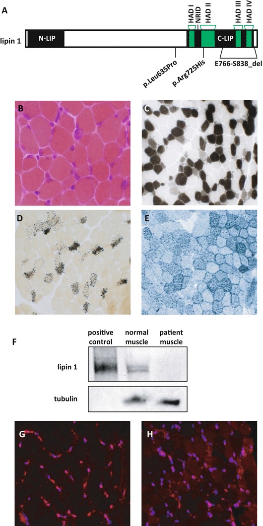
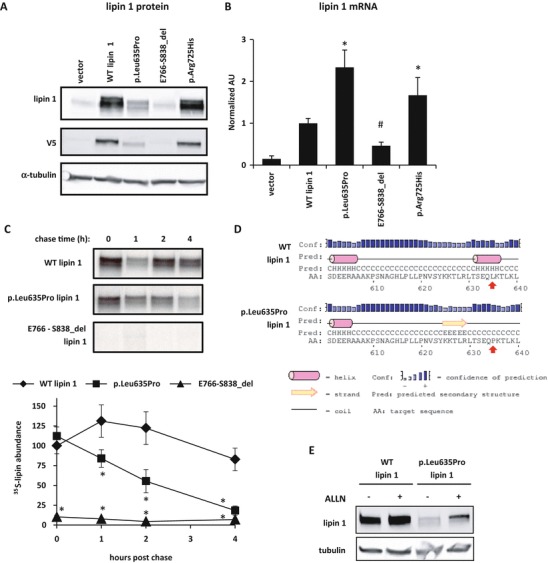
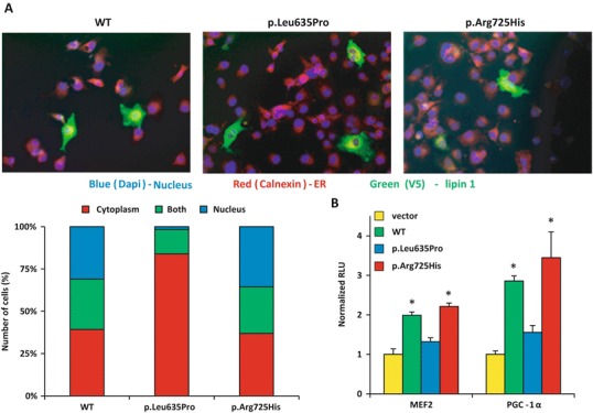
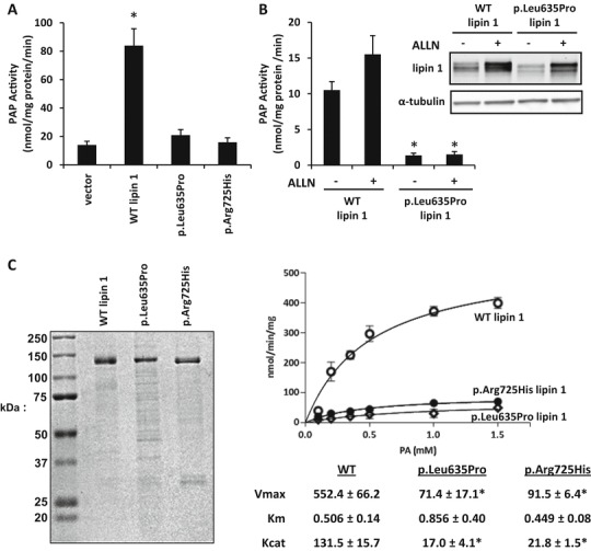
Rhabdomyolysis is an acute syndrome due to extensive injury of skeletal muscle. Recurrent rhabdomyolysis is often caused by inborn errors in intermediary metabolism, and recent work has suggested that mutations in the human gene encoding lipin 1 (LPIN1) may be a common cause of recurrent rhabdomyolysis in children. Lipin 1 dephosphorylates phosphatidic acid to form diacylglycerol (phosphatidic acid phosphohydrolase; PAP) and acts as a transcriptional regulatory protein to control metabolic gene expression. Herein, a 3-year-old boy with severe recurrent rhabdomyolysis was determined to be a compound heterozygote for a novel c.1904T>C (p.Leu635Pro) substitution and a previously reported genomic deletion of exons 18-19 (E766-S838_del) in LPIN1. Western blotting with patient muscle biopsy lysates demonstrated a marked reduction in lipin 1 protein, while immunohistochemical staining for lipin 1 showed abnormal subcellular localization. We cloned cDNAs to express recombinant lipin 1 proteins harboring pathogenic mutations and showed that the E766-S838_del allele was not expressed at the RNA or protein level. Lipin 1 p.Leu635Pro was expressed, but the protein was less stable, was aggregated in the cytosol, and was targeted for proteosomal degradation. Another pathogenic single amino acid substitution, lipin 1 p.Arg725His, was well expressed and retained its transcriptional regulatory function. However, both p.Leu635Pro and p.Arg725His proteins were found to be deficient in PAP activity. Kinetic analyses demonstrated a loss of catalysis rather than diminished substrate binding. These data suggest that loss of lipin 1-mediated PAP activity may be involved in the pathogenesis of rhabdomyolysis in lipin 1 deficiency.


Figures

Fig. 1
Fig. 1
LPIN1 mutations in a boy with recurrent rhabdomyolysis. (a) Diagram of LPIN1 gene. The haloacid dehalogenase (HAD) domains that are important for regulating PAP activity are noted. The nuclear receptor interaction domain (NRID) is important for transcriptional regulatory function. Locations of LPIN1 mutations of the patient (p.Leu635Pro and E766-S838_del) are included with the location of another reported mutation (p.Arg725His). (bh) A muscle biopsy was performed 6 weeks after the patient’s second episode of rhabdomyolysis and processed for routine histochemical staining. All photomicrographs were obtained at 20× magnification. (b) H&E demonstrated moderate variation in fiber size with occasional basophilic fibers; (c) ATPase 4.3 revealed an excessive number of Type IIC muscle fibers; (d) alkaline phosphatase with increased number of positive staining fibers; (e) Sudan black with moderately increased lipid. (f) Western blot demonstrating near absence of lipin 1 expression in the same patient. (g) Lipin 1 immunostaining in a pediatric control muscle shows strong nuclear staining of lipin 1. (h) Lipin 1 immunostaining in the p.Leu635Pro; E766-S838_del compound heterozygote muscle shows significantly reduced lipin 1 staining with an abnormal stippled or perinuclear staining pattern for the small amount of protein that is present (Blue: DAPI, Red: lipin 1)
Fig. 2
Fig. 2
Lipin 1 proteins with disease-associated mutations have reduced protein abundance despite increased RNA levels and reduced protein stability. (a) Representative western blots using lysates from HEK293 cells transfected with V5-tagged lipin 1 (wild-type or mutant) or vector-only control are shown using antibodies against lipin 1, V5, and α-tubulin. (b) Lipin 1 mRNA expression of HEK293 cells transfected with V5-tagged lipin 1 (wild type or mutant) or vector only. Compared to WT lipin 1, p.Leu635Pro and p.Arg725His have significantly greater (*P < 0.05, t-test) and E766-S838 has significantly lower mRNA (#P < 0.05, t-test) expression. (c) A representative autoradiograph and average percent of radiolabeled lipin 1 abundance relative to WT lipin 1 from pulse-chase studies calculated from three separate gels are shown. Protein half-life of p.Leu635Pro was significantly lower than WT lipin 1 at 1, 2, and 4 h (*P < 0.05, t-test). (d) The panel depicts the results of PSIPRED prediction of the secondary structures of the affected region of lipin 1 proteins. The red arrows indicate the affected amino acids. p.Leu635Pro mutation results in disruption of a predicted α-helix and possible formation of a β-strand. (e) Representative western blots of HEK293 cells with overexpressed V5-tagged lipin 1 and p.Leu635Pro are shown. Cells were treated for 4 h with 26 μM ALLN prior to harvest
Fig. 3
Fig. 3
Nuclear localization and transcriptional regulatory function of lipin 1 mutant proteins. (a) COS7 cells transfected with V5-tagged lipin 1 (wild type or mutant) to examine its subcellular localization with the nuclear marker (DAPI) and the endoplasmic reticulum marker (calnexin). The percentage of the number of cells analyzed that localized to the cytoplasm, nucleus, or both was measured in at least 60 cells in at least 15 distinct fields. L365P had very little nuclear localization compared to WT and p.Arg725His. (b) Coactivation of Gal4-MEF2A and PGC-1α in COS7 cells cotransfected with a Gal4-responsive UAS-TK-luciferase reporter construct and cDNAs to express lipin 1 (WT or p.Arg725His), Gal4-MEF2A, Gal4-PGC-1α, or vector control. *p < 0.05 vs. vector control
Fig. 4
Fig. 4
Lipin 1 proteins with disease-associated mutations lack PAP activity. (a) PAP activity of HEK293 cells overexpressing lipin 1 and lipin 1 mutants. PAP activity of WT lipin 1 was significantly greater than vector, p.Leu635Pro, and p.Arg725His (*P < 0.05, t-test). (b) PAP activity of HEK293 cells overexpressing lipin 1 and p.Leu635Pro incubated with ALLN. PAP activity of untreated WT lipin 1 was significantly greater than ALLN-treated and ALLN-untreated p.Leu635Pro (*P < 0.05, t-test). (c) Coomassie stain and PAP activity of purified recombinant WT lipin 1, p.Leu635Pro, and p.Arg725His. PAP activity was measured using Triton X-100/PA mixed micelles at pH 7.5. WT lipin 1 had approx. sixfold higher V max than p.Leu635Pro and p.Arg725His with no change in K m

References

    1. Bennett MJ. Pathophysiology of fatty acid oxidation disorders. J Inherit Metab Dis. 2010;33:533–537. doi: 10.1007/s10545-010-9170-y. - DOI - PubMed
    1. Buchan DW, Minneci F, Nugent TC, Bryson K, Jones DT. Scalable web services for the PSIPRED protein analysis workbench. Nucleic Acids Res. 2013;41:W349–W357. doi: 10.1093/nar/gkt381. - DOI - PMC - PubMed
    1. Castets P, Lin S, Rion N, et al. Sustained activation of mTORC1 in skeletal muscle inhibits constitutive and starvation-induced autophagy and causes a severe, late-onset myopathy. Cell Metab. 2013;17:731–744. doi: 10.1016/j.cmet.2013.03.015. - DOI - PubMed
    1. Eaton JM, Mullins GR, Brindley DN, Harris TE. Phosphorylation of lipin 1 and charge on the phosphatidic acid head group control its phosphatidic acid phosphatase activity and membrane association. J Biol Chem. 2013;288:9933–9945. doi: 10.1074/jbc.M112.441493. - DOI - PMC - PubMed
    1. Finck BN, Gropler MC, Chen Z, et al. Lipin 1 is an inducible amplifier of the hepatic PGC-1alpha/PPARalpha regulatory pathway. Cell Metab. 2006;4:199–210. doi: 10.1016/j.cmet.2006.08.005. - DOI - PubMed