. 2015 Jul 15;469(2):223-33.
doi: 10.1042/BJ20150396. Epub 2015 May 13.

Nuclear cyclophilins affect spliceosome assembly and function in vitro

Affiliations

Nuclear cyclophilins affect spliceosome assembly and function in vitro

B M Adams et al. Biochem J. .

Abstract

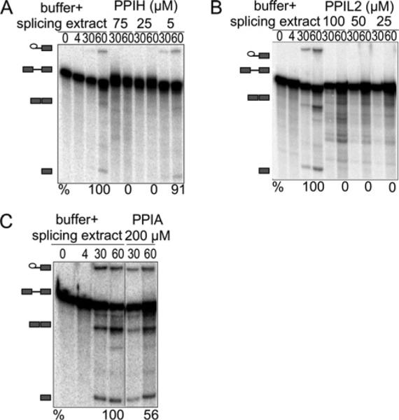
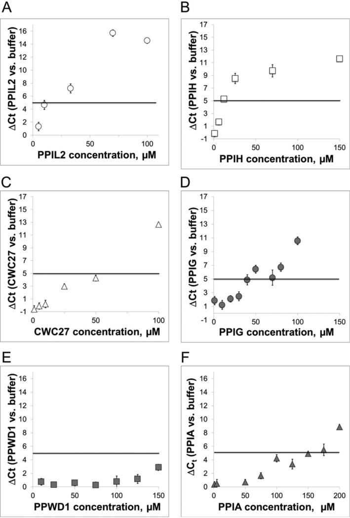
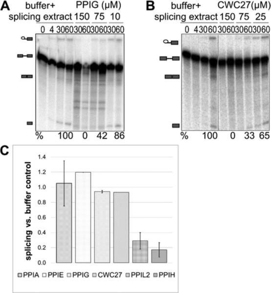
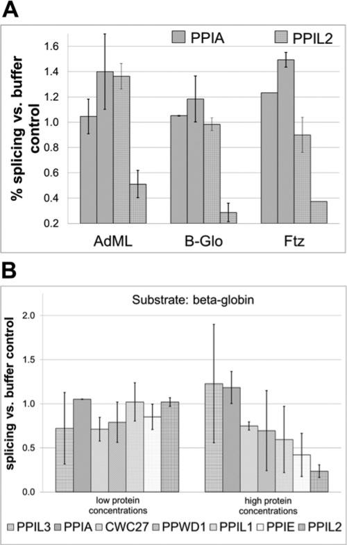
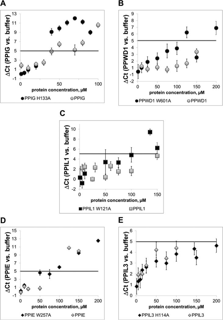
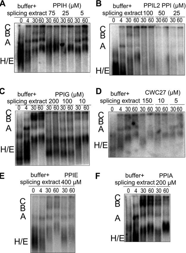
Cyclophilins are ubiquitously expressed proteins that bind to prolines and can catalyse cis/trans isomerization of proline residues. There are 17 annotated members of the cyclophilin family in humans, ubiquitously expressed and localized variously to the cytoplasm, nucleus or mitochondria. Surprisingly, all eight of the nuclear localized cyclophilins are found associated with spliceosomal complexes. However, their particular functions within this context are unknown. We have therefore adapted three established assays for in vitro pre-mRNA splicing to probe the functional roles of nuclear cyclophilins in the context of the human spliceosome. We find that four of the eight spliceosom-associated cyclophilins exert strong effects on splicing in vitro. These effects are dose-dependent and, remarkably, uniquely characteristic of each cyclophilin. Using both qualitative and quantitative means, we show that at least half of the nuclear cyclophilins can act as regulatory factors of spliceosome function in vitro. The present work provides the first quantifiable evidence that nuclear cyclophilins are splicing factors and provides a novel approach for future work into small molecule-based modulation of pre-mRNA splicing.

Keywords: nuclear cyclophilins; pre-messenger RNA (mRNA) splicing; spliceosome.


Figures

Figure 1
Figure 1. PPIH and PPIL2 inhibit splicing in vitro as measured by denaturing gel electrophoresis
The panels show denaturing gel analysis of radiolabelled HMS388 splicing substrate, isolated from in vitro splicing reactions supplemented with increasing concentrations of the indicated cyclophilin proteins or with buffer alone. RNA species are schematized at the left as (from top to bottom) lariat intron intermediate, pre-mRNA, mRNA, free 5′-exon. Percentage splicing efficiency (amount of mRNA compared with total RNA species) is indicated at the bottom of each gel image. As indicated, relatively low concentrations of PPIH (A) or PPIL2 (B) efficiently inhibit the splicing reaction; in contrast, high concentrations of PPIA (C) do not block splicing activity. Note that unless indicated by a black line, lanes are contiguous and from a single gel, although experiments were performed at least three times. Also note that identical splicing substrate (HMS388) is loaded in each lane; slight differences in mobility are probably due to effects from overloading samples in order to detect low populations of splicing intermediates.
Figure 2
Figure 2. PPIL2, PPIH, CWC27 and PPIG inhibit splicing as measured by an exon-junction specific probe
The amount of AdML mRNA produced in the splicing reaction is detected via RT-qPCR by a TaqMan probe to the spliced exon junction sequence. Plotted is the average difference in cycles required to reach threshold (ΔCt) between reactions supplemented with cyclophilins compared with buffer alone. The five-cycle ΔCt threshold, as mentioned in the text, corresponds to significant inhibition in the production of mRNA and is shown in all RT-qPCR plots as a solid black line. We find that PPIL2 (A) and PPIH (B) are strong inhibitors of our AdML splicing substrate; CWC27 (C) and PPIG (D) slightly less so; and the other four nuclear cyclophilins are not inhibitory in this experimental setup [see PPWD1 in panel (E) and also Supplementary Figure S1]. The control PPIA (F), as in Figure 1, shows only slight inhibition of splicing at high protein concentrations.
Figure 3
Figure 3. Inhibition of splicing as seen in the RT-qPCR assay is replicated using denaturing gel analysis
(A) PPIG is shown to inhibit splicing of HMS388 splicing substrate. (B) CWC27 is also shown to be inhibitory. Panels are labelled as in Figure 1. Panel (C) shows quantification of splicing efficiency for PPIA, PPIE, PPIG, CWC27, PPIL2 and PPIH using denaturing gel analysis. Values and associated error are calculated by averaging the data from multiple lanes on acrylamide gels.
Figure 4
Figure 4. The nuclear cyclophilins inhibit splicing of multiple pre-mRNA substrates
(A) A low and a high concentration of PPIA and PPIL2 are tested against the AdML, β-globin and ftz substrates. Results are quantified from denaturing gel analysis and error calculated as the S.D. between lanes on the gel. (B) Results for six nuclear cyclophilins and the control PPIA are quantified for the β-globin substrate. In general, the nuclear cyclophilins characterized as inhibitory using the AdML substrate are also inhibitory against β-globin. PPIL3 and PPIA are slightly stimulatory and PPWD1 and PPIL1 are inhibitory against β-globin.
Figure 5
Figure 5. The isomerase activity of nuclear cyclophilins is not necessary to detect splicing inhibition
For cyclophilins that are active isomerases against proline-containing substrates, point mutations were made at the position corresponding to Trp121 in PPIA. This mutation should abrogate proline turnover and cyclosporine binding [39]. In panels (A) and (B), it can be seen that mutation of PPIG or PPWD1 increases splicing inhibition. In panel (C), the modest effect of mutating PPIL1 is seen. In (D and E), mutation of the inhibitory PPIE PPI domain or of PPIL3 has no effect on splicing inhibition.
Figure 6
Figure 6. The effect of cyclophilins on spliceosome assembly visualized by native agarose gel analysis of in vitro splicing reactions
In each panel, the first four lanes show a time course of splicing reactions with buffer and nuclear extract, along with the radiolabelled AdML substrate. The relative positions of E/H, A, B and C complexes are indicated to the left of each gel. Subsequent lanes show 30 and 60 min time-points of splicing reactions containing the indicated concentrations of PPIH (A), PPIL2 (B), PPIG (C), CWC27 (D), PPIE (E) or PPIA (F).

References

    1. Valadkhan S, Jaladat Y. The spliceosomal proteome: at the heart of the largest cellular ribonucleoprotein machine. Proteomics. 2010;10:4128–4141. CrossRef PubMed. - PMC - PubMed
    1. Wahl MC, Will CL, Lührmann R. The spliceosome: design principles of a dynamic RNP machine. Cell. 2009;136:701–718. CrossRef PubMed. - PubMed
    1. Will CL, Lührmann R. Spliceosome structure and function. Cold Spring Harb. Perspect. Biol. 2011;3:a0037074. CrossRef. - PMC - PubMed
    1. Jurica MS, Licklider LJ, Gygi SR, Grigorieff N, Moore MJ. Purification and characterization of native spliceosomes suitable for three-dimensional structural analysis. RNA. 2002;8:426–439. CrossRef PubMed. - PMC - PubMed
    1. Agafonov DE, Deckert J, Wolf E, Odenwalder P, Bessonov S, Will CL, Urlaub H, Luhrmann R. Semiquantitative proteomic analysis of the human spliceosome via a novel two-dimensional gel electrophoresis method. Mol. Cell. Biol. 2011;13:2667–2682. CrossRef. - PMC - PubMed

Publication types