

The Effect of Lesion Size on the Organization of the Ipsilesional and Contralesional Motor Cortex
- PMID: 25967757
- PMCID: PMC4766967
- DOI: 10.1177/1545968315585356
The Effect of Lesion Size on the Organization of the Ipsilesional and Contralesional Motor Cortex
Abstract
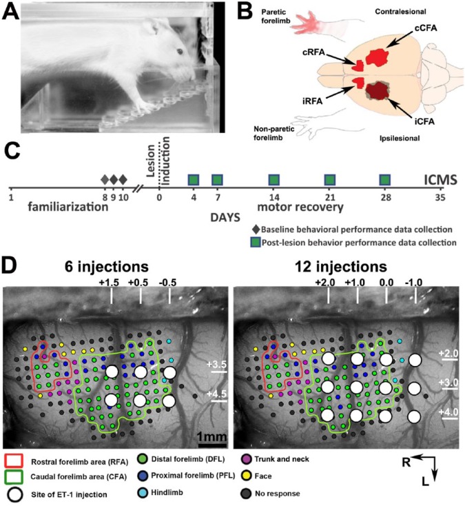
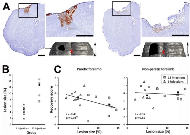
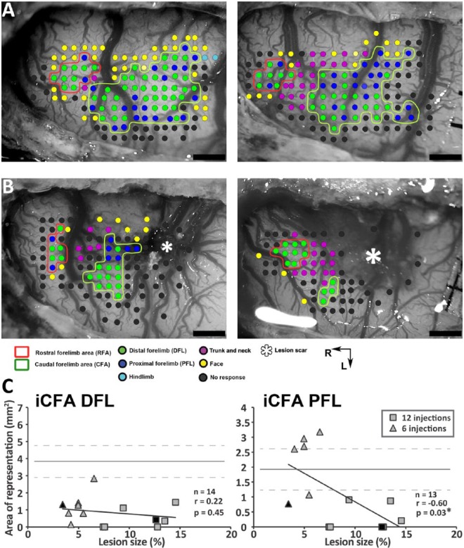
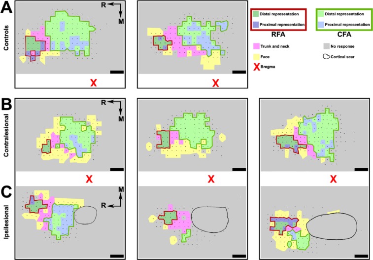
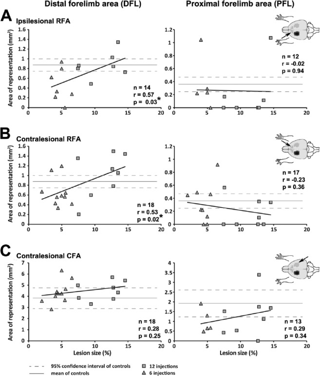
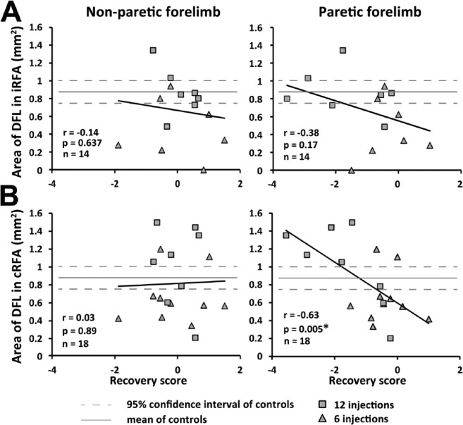
Recovery of hand function following lesions in the primary motor cortex (M1) is associated with a reorganization of premotor areas in the ipsilesional hemisphere, and this reorganization depends on the size of the lesion. It is not clear how lesion size affects motor representations in the contralesional hemisphere and how the effects in the 2 hemispheres compare. Our goal was to study how lesion size affects motor representations in the ipsilesional and contralesional hemispheres. In rats, we induced lesions of different sizes in the caudal forelimb area (CFA), the equivalent of M1. The effective lesion volume in each animal was quantified histologically. Behavioral recovery was evaluated with the Montoya Staircase task for 28 days after the lesion. Then, the organization of the CFA and the rostral forelimb area (RFA)--the putative premotor area in rats--in the 2 cerebral hemispheres was studied with intracortical microstimulation mapping techniques. The distal forelimb representation in the RFA of both the ipsilesional and contralesional hemispheres was positively correlated with the size of the lesion. In contrast, lesion size had no effect on the contralesional CFA, and there was no relationship between movement representations in the 2 hemispheres. Finally, only the contralesional RFA was negatively correlated with chronic motor deficits of the paretic forelimb. Our data show that lesion size has comparable effects on motor representations in premotor areas of both hemispheres and suggest that the contralesional premotor cortex may play a greater role in the recovery of the paretic forelimb following large lesions.
Keywords: contralesional; cortex; hand; intact hemisphere; ipsilesional; lesion size; motor representations; plasticity; recovery; stroke.
© The Author(s) 2015.
Conflict of interest statement
Figures
Similar articles
-
Neural network remodeling underlying motor map reorganization induced by rehabilitative training after ischemic stroke.Neuroscience. 2016 Dec 17;339:338-362. doi: 10.1016/j.neuroscience.2016.10.008. Epub 2016 Oct 8. Neuroscience. 2016. PMID: 27725217
-
Traumatic Brain Injury Occludes Training-Dependent Cortical Reorganization in the Contralesional Hemisphere.J Neurotrauma. 2017 Sep;34(17):2495-2503. doi: 10.1089/neu.2016.4796. Epub 2017 Jul 19. J Neurotrauma. 2017. PMID: 28462608 Free PMC article.
-
Plasticity in One Hemisphere, Control From Two: Adaptation in Descending Motor Pathways After Unilateral Corticospinal Injury in Neonatal Rats.Front Neural Circuits. 2018 Apr 12;12:28. doi: 10.3389/fncir.2018.00028. eCollection 2018. Front Neural Circuits. 2018. PMID: 29706871 Free PMC article.
-
The role of the contralesional primary motor cortex in upper limb recovery after stroke: a scoping review following PRISMA-ScR guidelines.BMC Neurosci. 2025 May 25;26(1):31. doi: 10.1186/s12868-025-00950-y. BMC Neurosci. 2025. PMID: 40414854 Free PMC article.
-
Inhibition of the contralesional hemisphere after stroke: reviewing a few of the building blocks with a focus on animal models.Prog Brain Res. 2015;218:361-87. doi: 10.1016/bs.pbr.2015.01.002. Epub 2015 Mar 30. Prog Brain Res. 2015. PMID: 25890146 Review.
Cited by
-
The Duration of Motor Responses Evoked with Intracortical Microstimulation in Rats Is Primarily Modulated by Stimulus Amplitude and Train Duration.PLoS One. 2016 Jul 21;11(7):e0159441. doi: 10.1371/journal.pone.0159441. eCollection 2016. PLoS One. 2016. PMID: 27442588 Free PMC article.
-
Facilitating Corticomotor Excitability of the Contralesional Hemisphere Using Non-Invasive Brain Stimulation to Improve Upper Limb Motor Recovery from Stroke-A Scoping Review.J Clin Med. 2024 Jul 28;13(15):4420. doi: 10.3390/jcm13154420. J Clin Med. 2024. PMID: 39124687 Free PMC article.
-
Inosine enhances recovery of grasp following cortical injury to the primary motor cortex of the rhesus monkey.Restor Neurol Neurosci. 2016 Sep 21;34(5):827-48. doi: 10.3233/RNN-160661. Restor Neurol Neurosci. 2016. PMID: 27497459 Free PMC article.
-
Evaluating the Abnormality of Bilateral Motor Cortex Activity in Subacute Stroke Patients Executing a Unimanual Motor Task With Increasing Demand on Precision.Front Neurol. 2022 May 25;13:836716. doi: 10.3389/fneur.2022.836716. eCollection 2022. Front Neurol. 2022. PMID: 35693005 Free PMC article.
-
Visual feedback alters force control and functional activity in the visuomotor network after stroke.Neuroimage Clin. 2017 Nov 14;17:505-517. doi: 10.1016/j.nicl.2017.11.012. eCollection 2018. Neuroimage Clin. 2017. PMID: 29201639 Free PMC article. Clinical Trial.
References
-
- Dancause N. Vicarious function of remote cortex following stroke: recent evidence from human and animal studies. Neuroscientist. 2006;12:489-499. - PubMed
-
- Nudo RJ. Mechanisms for recovery of motor function following cortical damage. Curr Opin Neurobiol. 2006;16:638-644. - PubMed
-
- Dancause N, Barbay S, Frost SB, et al. Effects of small ischemic lesions in the primary motor cortex on neurophysiological organization in ventral premotor cortex. J Neurophysiol. 2006;96:3506-3511. - PubMed
-
- Frost SB, Barbay S, Friel KM, Plautz EJ, Nudo RJ. Reorganization of remote cortical regions after ischemic brain injury: a potential substrate for stroke recovery. J Neurophysiol. 2003;89:3205-3214. - PubMed
Publication types
MeSH terms
Substances
Grants and funding
LinkOut - more resources
Full Text Sources
Other Literature Sources
