Review
. 2015 Oct;88(1054):20150207.
doi: 10.1259/bjr.20150207. Epub 2015 Jun 12.

Applications of nanoparticles for diagnosis and therapy of cancer

Affiliations
Review

Applications of nanoparticles for diagnosis and therapy of cancer

S C Baetke et al. Br J Radiol. 2015 Oct.

Abstract

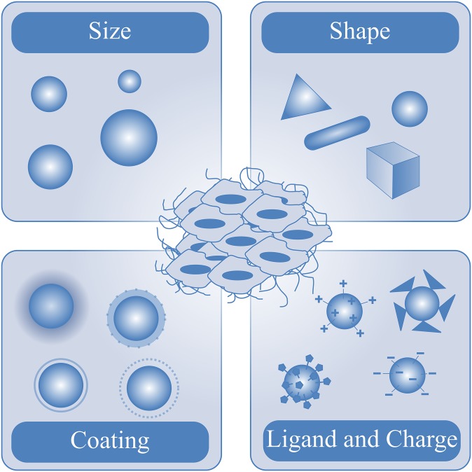
During the last decades, a plethora of nanoparticles have been developed and evaluated and a real hype has been created around their potential application as diagnostic and therapeutic agents. Despite their suggestion as potential diagnostic agents, only a single diagnostic nanoparticle formulation, namely iron oxide nanoparticles, has found its way into clinical routine so far. This fact is primarily due to difficulties in achieving appropriate pharmacokinetic properties and a reproducible synthesis of monodispersed nanoparticles. Furthermore, concerns exist about their biodegradation, elimination and toxicity. The majority of nanoparticle formulations that are currently routinely used in the clinic are used for therapeutic purposes. These therapeutic nanoparticles aim to more efficiently deliver a (chemo-) therapeutic drug to the pathological site, while avoiding its accumulation in healthy organs and tissues, and are predominantly based on the "enhanced permeability and retention" (EPR) effect. Furthermore, based on their ability to integrate diagnostic and therapeutic entities within a single nanoparticle formulation, nanoparticles hold great promise for theranostic purposes and are considered to be highly useful for personalizing nanomedicine-based treatments. In this review article, we present applications of diagnostic and therapeutic nanoparticles, summarize frequently used non-invasive imaging techniques and describe the role of EPR in the accumulation of nanotheranostic formulations. In this context, the clinical potential of nanotheranostics and image-guided drug delivery for individualized and improved (chemo-) therapeutic interventions is addressed.


Figures

Figure 1.
Figure 1.
Main properties of nanoparticles influencing their biodistribution, elimination and target site accumulation.
Figure 2.
Figure 2.
Characterization of tumour vascularization and angiogenesis by particulate contrast agents. (a) Molecular ultrasound imaging of unconjugated, RGD-conjugated microbubbles targeted against αvβ3 integrin and VEGFR2-conjugated microbubbles, demonstrating specific binding of RGD- and VEGFR2-conjugated microbubbles to angiogenic tumour blood vessels. (b) MR molecular imaging of RGD-conjugated and RAD-modified control liposomal nanoparticles in tumour-bearing mice, showing differences in the accumulation pattern of the RGD-conjugated and RAD-modified control liposomes, which closely correlates with the position of angiogenic blood vessels in the tumour. (c) MR images of HaCaT-ras A-5RT3 tumours before and 6 h after intravenous (i.v.) injection of RGD-USPIO (ultrasmall superparamagnetic iron oxide) and USPIO nanoparticles, showing focal areas with a strong and heterogeneous decrease in signal intensity after injection of RGD-USPIO nanoparticles. (d) Fluorescence reflectance imaging of tumour accumulation of an RGD-based polymeric nanocarrier (P-RGD) and a control copolymer (P-CON) in CT26 tumours 1 and 72 h after i.v. injection, showing early binding of the actively targeted probe P-RGD to tumour blood vessels, while the passively targeted probe P-CON showed progressive enhanced permeability and retention (EPR)-mediated tumour accumulation. (e) Positron emission tomography (PET)-CT imaging of the biodistribution and tumour uptake of 124I-cRGDY-PEG-C-dots in a patient with anorectal mucosal melanoma and known liver metastasis in the left hepatic lobe. (I) Coronal CT image showing a hypodense liver metastasis. (II) PET and (III) co-registered PET-CT imaging 4 h after i.v. administration of the nanoparticles demonstrate nanoparticle uptake, which appeared to be restricted to the tumour margin, as well as nanoparticle activity in the gastrointestinal tract, gallbladder, bladder and heart. (IV) PET-CT imaging 24 h after nanoparticle administration revealed clearance of the nanoparticle activity with some remaining particle activity in the tumour margin. (V) A corresponding 18F-FDG PET-CT scan acquired several days after PET-CT imaging validated the localization of the liver metastasis. Adapted from Zhang et al, Phillips et al (with permission from the American Association for the Advancement of Science), Palmowski et al, Mulder et al (with permission from the Federation of American Societies for Experimental Biology) and Kunjachan et al (with permission from the American Chemical Society).
Figure 3.
Figure 3.
Theranostic applications of nanoparticles. (a) Scintigraphic analysis of the biodistribution of two differently sized 131I-labelled HPMA copolymers in AT1 tumours, demonstrating effective tumour accumulation and prolonged circulation (B, bladder; H, heart; L, liver; S, spleen; T, tumour). (b) Tumour and organ concentrations of the two radiolabeled copolymers 24 and 168 h after intravenous (i.v.) administration, showing significantly higher concentrations in tumour compared with the concentrations in other healthy organs (except for the lung and spleen). (c) MicroSPECT-CT imaging (left panel) and scintigraphic analysis (right panel) of 186Re-labelled Doxil® in HNSCC-bearing nude rats 20 h after i.v. administration of 186Re-labelled Doxil, demonstrating prolonged blood retention and effective tumour accumulation of 186Re-labelled Doxil (H, heart; L, liver; S, spleen; T, tumour). (d) Planar scintigraphic analysis of stealth liposomal doxorubicin in patients suffering from sarcomas undergoing radiotherapy reveals intense drug accumulation in tumours, demonstrating the feasibility of visualizing and quantifying EPR-mediated passive drug targeting to tumours (from left to right: fibrosarcoma of the iliac region, angiosarcoma of the maxillary antrum, Ewing sarcoma of the femur, Kaposi sarcoma of the palmar region). Adapted from Koukourakis et al, Lammers et al and Soundararajan et al with permission from Informa Healthcare, Nature Publishing Group and Elsevier, respectively.
Figure 4.
Figure 4.
Concepts for theranostics and personalized medicine. Such strategies include patient screening, diagnosis, treatment and follow-up monitoring, and are based on “companion diagnostics” and “image-guidance.” The application of companion diagnostics allows patient preselection based on patient profiling of specific biomarkers or gene signatures and enables therapy selection and treatment response prediction. Image-guided drug delivery of theranostic nanoparticles enables monitoring of their biodistribution and target site accumulation, the visualization and quantification of their local activation and sometimes even drug release and the non-invasive and longitudinal assessment of their therapeutic efficacy.

Similar articles

Cited by

References

    1. Bao G, Mitragotri S, Tong S. Multifunctional nanoparticles for drug delivery and molecular imaging. Annu Rev Biomed Eng 2013; 15: 253–82. doi: 10.1146/annurev-bioeng-071812-152409 - DOI - PMC - PubMed
    1. Cormode DP, Skajaa T, Fayad ZA, Mulder WJ. Nanotechnology in medical imaging: probe design and applications. Arterioscler Thromb Vasc Biol 2009; 29: 992–1000. doi: 10.1161/ATVBAHA.108.165506 - DOI - PMC - PubMed
    1. Key J, Leary JF. Nanoparticles for multimodal in vivo imaging in nanomedicine. Int J Nanomedicine 2014; 9: 711–26. doi: 10.2147/IJN.S53717 - DOI - PMC - PubMed
    1. Chen ZY, Wang YX, Lin Y, Zhang JS, Yang F, Zhou QL, et al. . Advance of molecular imaging technology and targeted imaging agent in imaging and therapy. Biomed Res Int 2014; 2014: 819324. doi: 10.1155/2014/819324 - DOI - PMC - PubMed
    1. Weissleder R, Pittet MJ. Imaging in the era of molecular oncology. Nature 2008; 452: 580–9. doi: 10.1038/nature06917 - DOI - PMC - PubMed

Publication types

MeSH terms

Substances