

Cavum Septum Pellucidum in Retired American Pro-Football Players
- PMID: 25970145
- PMCID: PMC4696427
- DOI: 10.1089/neu.2014.3805
Cavum Septum Pellucidum in Retired American Pro-Football Players
Abstract
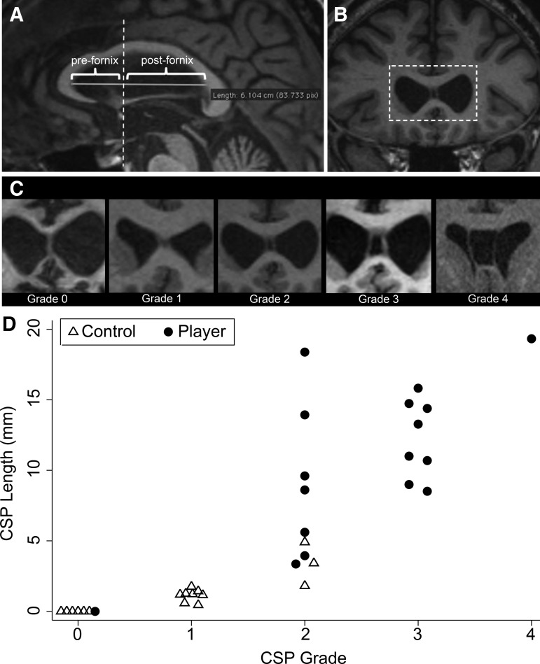
Previous studies report that cavum septum pellucidum (CSP) is frequent among athletes with a history of repeated traumatic brain injury (TBI), such as boxers. Few studies of CSP in athletes, however, have assessed detailed features of the septum pellucidum in a case-control fashion. This is important because prevalence of CSP in the general population varies widely (2% to 85%) between studies. Further, rates of CSP among American pro-football players have not been described previously. We sought to characterize MRI features of the septum pellucidum in a series of retired pro-football players with a history of repeated concussive/subconcussive head traumas compared with controls. We retrospectively assessed retired American pro-football players presenting to our memory clinic with cognitive/behavioral symptoms in whom structural MRI was available with slice thickness ≤2 mm (n=17). Each player was matched to a memory clinic control patient with no history of TBI. Scans were interpreted by raters blinded to clinical information and TBI/football history, who measured CSP grade (0-absent, 1-equivocal, 2-mild, 3-moderate, 4-severe) and length according to a standard protocol. Sixteen of 17 (94%) players had a CSP graded ≥2 compared with 3 of 17 (18%) controls. CSP was significantly higher grade (p<0.001) and longer in players than controls (mean length±standard deviation: 10.6 mm±5.4 vs. 1.1 mm±1.3, p<0.001). Among patients presenting to a memory clinic, long high-grade CSP was more frequent in retired pro-football players compared with patients without a history of TBI.
Keywords: concussion; magnetic resonance imaging; septum pellucidum; traumatic brain injury.
Figures
References
-
- McKee A.C., Stern R.A., Nowinski C.J., Stein T.D., Alvarez V.E., Daneshvar D.H., Lee H.S., Wojtowicz S.M., Hall G., Baugh C.M., Riley D.O., Kubilus C.A., Cormier K.A., Jacobs M.A., Martin B.R., Abraham C.R., Ikezu T., Reichard R.R., Wolozin B.L., Budson A.E., Goldstein L.E., Kowall N.W., and Cantu R.C. (2013). The spectrum of disease in chronic traumatic encephalopathy. Brain 136, 43–64 - PMC - PubMed
-
- Cabanis E.A., Perez G., Tamraz J.C., Iba-Zizen M.T., Roger B., Alfonso J.M., and Rougemont D. (1986). Cephalic magnetic resonance imaging of boxers. Preliminary results. Acta Radiol. Suppl. 369, 365–366 - PubMed
-
- Orrison W.W., Hanson E.H., Alamo T., Watson D., Sharma M., Perkins T.G. and Tandy R.D. (2009). Traumatic brain injury: a review and high-field MRI findings in 100 unarmed combatants using a literature-based checklist approach. J. Neurotrauma 26, 689–701 - PubMed
-
- Bogdanoff B., and Natter H.M. (1989). Incidence of cavum septum pellucidum in adults: a sign of boxer's encephalopathy. Neurology 39, 991–992 - PubMed
Publication types
MeSH terms
Grants and funding
LinkOut - more resources
Full Text Sources
Other Literature Sources
Medical
