. 2015 Jul 2;523(7558):101-5.
doi: 10.1038/nature14357. Epub 2015 May 11.

Cytosolic extensions directly regulate a rhomboid protease by modulating substrate gating

Affiliations

Cytosolic extensions directly regulate a rhomboid protease by modulating substrate gating

Rosanna P Baker et al. Nature. .

Abstract

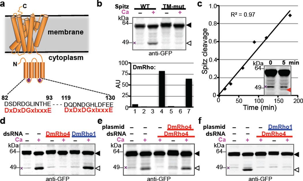
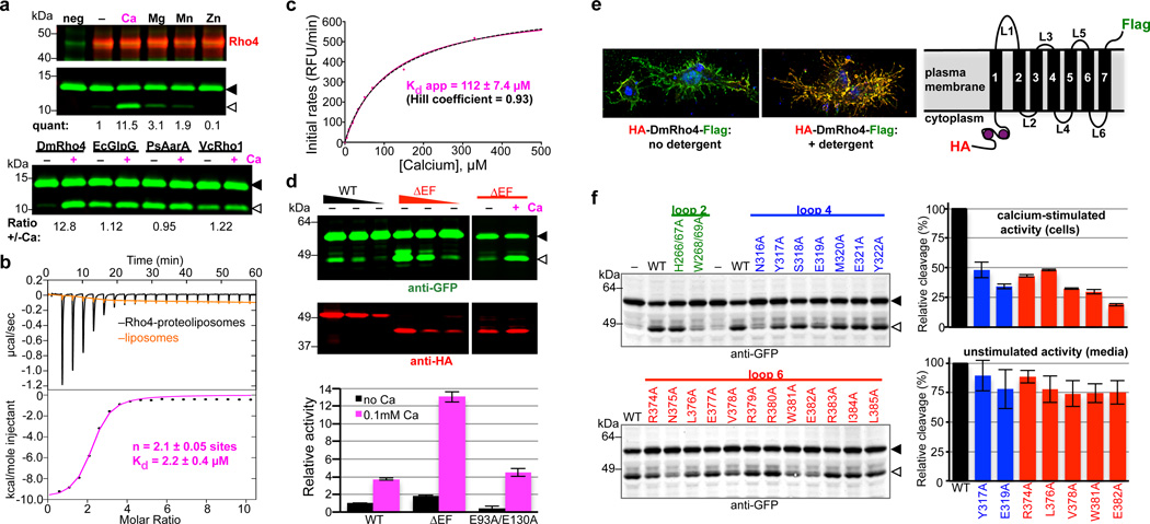
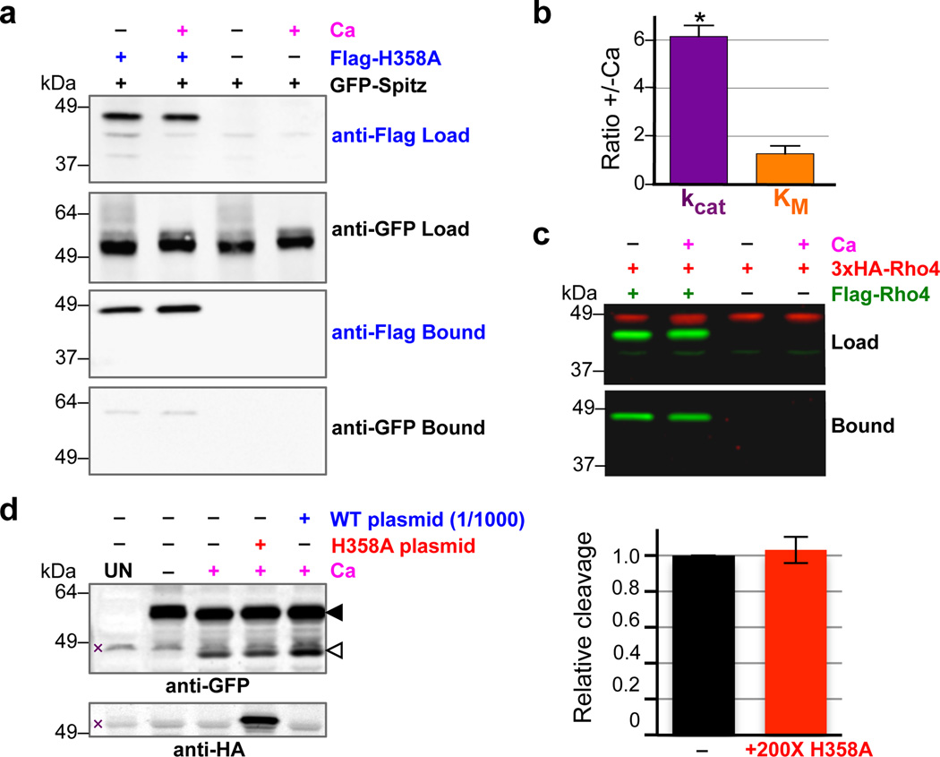
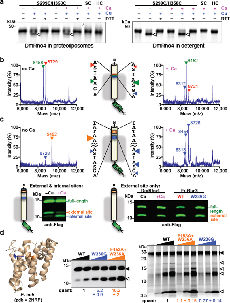
Intramembrane proteases catalyse the signal-generating step of various cell signalling pathways, and continue to be implicated in diseases ranging from malaria infection to Parkinsonian neurodegeneration. Despite playing such decisive roles, it remains unclear whether or how these membrane-immersed enzymes might be regulated directly. To address this limitation, here we focus on intramembrane proteases containing domains known to exert regulatory functions in other contexts, and characterize a rhomboid protease that harbours calcium-binding EF-hands. We find calcium potently stimulates proteolysis by endogenous rhomboid-4 in Drosophila cells, and, remarkably, when rhomboid-4 is purified and reconstituted in liposomes. Interestingly, deleting the amino-terminal EF-hands activates proteolysis prematurely, while residues in cytoplasmic loops connecting distal transmembrane segments mediate calcium stimulation. Rhomboid regulation is not orchestrated by either dimerization or substrate interactions. Instead, calcium increases catalytic rate by promoting substrate gating. Substrates with cleavage sites outside the membrane can be cleaved but lose the capacity to be regulated. These observations indicate substrate gating is not an essential step in catalysis, but instead evolved as a mechanism for regulating proteolysis inside the membrane. Moreover, these insights provide new approaches for studying rhomboid functions by investigating upstream inputs that trigger proteolysis.


Conflict of interest statement

The authors declare that no financial conflict of interest exists.

Figures

Extended Data Figure 1
Extended Data Figure 1. The Rhomboid-4 subfamily of rhomboid proteases
a ClustalW multiple sequence alignment of the conserved amino-terminal EF-Hand domains of 24 members of the Rhomboid-4 subfamily (generated in Biology Workbench, http://workbench.sdsc.edu). Identical residues are shaded in green, highly conserved residues in yellow and similar residues in cyan. The EF-hand calcium-binding loop consensus sequence is given below the alignment. b Rooted tree of the 24 Rhomboid-4 homologues. Genus species names are color-coded as follows: primates (blue), other mammals (green), birds (purple), fish (pink), non-vertebrate chordate (cyan), nematode (orange), and insects (red), with common names given in parenthesis, followed by NCBI accession numbers.
Extended Data Figure 2
Extended Data Figure 2. Activity and thermostability analysis of DmRho4 mutants
a Comparison of calcium stimulation of DmRho4 versus its EF hand domain deletion mutant (ΔEF), and a mutant lacking the entire cytosolic domain (ΔN). Upper diagram shows position of domains (demarked by residue numbers) and the corresponding deletion constructs. Transmembrane segments are shown as grey rectangles. GFP-Spitz substrate and cleavage products (green bands in the anti-GFP western) are denoted by black and white triangles, respectively. DmRho4 protein levels are shown as red bands (anti-HA western). b Analysis of DmRho4 loop 2, 4, and 6 mutant protein levels from Fig. 2f (calcium stimulation conditions). c DmRho4 loop 2, 4, and 6 mutants were assayed for cleavage of GFP-Spitz under basal (unstimulated) conditions for ~24 hours in the absence of calcium. Cleavage product (green bands, white arrowhead) was detected in media fractions for most of the mutants at levels comparable to the wild type enzyme. Corresponding DmRho4 protein levels are shown as red bands (anti-HA western analyses). d Wild type DmRho4 and engineered variants were expressed and purified from bacteria, subjected to quantitative thermal stability analysis, and transition temperature midpoints (Tms) were derived (error bars indicate the standard deviation of four experimental replicates). The thermal stability of mutant DmRho4 proteases was indistinguishable from that of wild type DmRho4.
Extended Data Figure 3
Extended Data Figure 3. Calcium does not regulate DmRho4 through intermolecular interactions
a Anti-Flag coimmunoprecipitation analysis of HA-DmRho4 and APP-Spi7-Flag substrate from proteoliposomes in the presence or absence of 0.5 mM calcium. An inactive mutant of DmRho4 (S299A) was used to facilitate substrate complex isolation. The amount of HA-tagged DmRo4 co-immunoprecipitated with the Flag-tagged substrate was not affected by the presence of 0.5mM calcium. L denotes ‘load’, B denotes ‘bound’. b Anti-Flag coimmunoprecipitation of Flag-DmRho4 and HA-DmRho4 from proteoliposomes. HA-tagged DmRho4 failed to coimmunoprecipitate with Flag-tagged DmRho4 in both the absence and presence of 0.5 mM calcium. c Mixing a catalytic mutant (S299A) and a calcium-binding mutant (E382A) cannot rescue calcium stimulation in trans (star indicates lane where a product would be expected with the mixed single mutants).
Extended Data Figure 4
Extended Data Figure 4. Lateral substrate gating underlies direct regulation of intramembrane proteolysis
a Thermostability analysis of single and double cysteine mutants of DmRho4 (error bars indicate the standard deviation of four experimental replicates). b Average relative proportions of cleavage at the external cleavage site (orange) compared to the internal cleavage site (blue) are shown for DmRho4 in the absence (no Ca) and presence (+ Ca) of 1 mM calcium (error bars indicate standard error of replicate experiments). The external site was favoured in the absence of calcium (approximately 80%) while internal cleavage was preferred in the presence of calcium (approximately 70%). c DmRho4 loop 4 and loop 6 calcium-binding site mutants retained calcium-independent cleavage of a substrate harbouring only an external cleavage site. Full-length substrate (solid triangle) and cleavage product (open triangle) are indicated. c Cleavage of a substrate with external and internal cleavage sites was compared for E. coli GlpG, P.stuartii AarA, and V. cholerae Rho1 in the absence (no Ca) or presence (+ Ca) of 0.5 mM calcium. The relative proportions of cleavage at the two sites varied between the bacterial rhomboid proteases, but in no case did calcium alter the cleavage site preference.
Figure 1
Figure 1. Calcium rapidly stimulates intramembrane proteolysis in Drosophila cells by endogenous DmRho4
a Diagram comparing the predicted calcium-binding loop residues of DmRho4 to an EF-hand consensus (in red). b Calcium ionophore treatment of Drosophila S2R+ cells induced cleavage of GFP-Spitz, but not its cleavage-site mutant, by endogenous DmRho4. Graph shows expression levels of Drosophila rhomboid genes in S2R+ cells (RNAseq data from modENCODE, modencode.org). c Ionophore-induced Spitz cleavage was detectable within 5 min (red triangle) and linear for 3h. d RNAi knockdown of DmRho4 but not of DmRho1 abrogated calcium-induced cleavage of GFP-Spitz. e Plasmid expression of DmRho4 rescued calcium-induced cleavage of GFP-Spitz in S2R+ cells undergoing RNAi. f Calcium-stimulated Spitz cleavage abolished by DmRho4 RNAi could not be rescued by DmRho1 overexpression. All images are anti-GFP western analyses, with substrate and cleavage bands denoted by black or open triangles, respectively, and non-specific bands marked by × (see Fig. 3d for untransfected cells).
Figure 2
Figure 2. Calcium directly regulates intramembrane proteolytic activity of DmRho4
a Proteolysis assay with pure reconstituted DmRho4 ± a panel of 1mM divalent metal ions (upper panels), and four different rhomboid enzymes reconstituted into proteoliposomes ± calcium (lower panel). EcGlpG is from Escherichia coli, PsAarA is from Providencia stuartii, and VcRho1 is from Vibrio cholerae. b Analysis of calcium binding to DmRho4 in proteoliposomes by isothermal titration calorimetry (upper graph shows the thermograms, lower graph is the liposome-subtracted quantification). c Calcium titration analysis of DmRho4 proteolysis using an inducible real-time reconstitution assay. Black dashed line shows an alternate fit with an optimal Hill coefficient. d Titration of wildtype (WT) and ∆EF DmRho4 in S2R+ cells comparing basal, unstimulated Spitz cleavage (left panel) and Spitz cleavage by DmRho4-∆EF ± calcium ionophore (right panel; also see Extended Data Set 2a). Lower graph shows in vitro activity of WT and EF-Hand mutants of DmRho4 in proteoliposomes (error bars indicate standard deviation for experimental replicates). e Topology of 3xHA-DmRho4-Flag in S2R+ cells as assessed by deconvolution immunofluorescence. The N-terminal HA-tag (red) was inaccessible while the C-terminal Flag tag (green) was accessible in the absence of detergent, indicating that the N-terminus is cytosolic while the C-terminus of DmRho4 is extracellular (blue marks nuclei). f Ability of DmRho4 cytosolic loop mutants to cleave GFP-Spitz in response to calcium ionophore stimulation in S2R+ cells was quantified by anti-GFP western analysis (also see Extended Data Set 2b for DmRho4 levels). Graphs show activity of selectively compromised loop 4 and 6 mutants under calcium-stimulated conditions in cells (upper graph) versus unstimulated conditions (lower graph, measured as cleavage product accumulation in culture media after 24 hours, also see Extended Data Fig. 2c). Error bars indicate standard deviation for experimental replicates. Black triangles and open triangles denote substrate and cleavage bands, respectively, throughout.
Figure 3
Figure 3. Intermolecular interactions do not mediate DmRho4 regulation by calcium
a Anti-Flag coimmunoprecipitation analysis of catalytically inactive Flag-DmRho4-H358A and GFP-Spitz from S2R+ cells untreated or treated with calcium ionophore. b Effect of calcium on the steady-state kinetic parameters of intramembrane proteolysis by reconstituted DmRho4 (mean ± standard deviation of 3 independent experiments, compared using a paired t-test). c Anti-Flag coimmunoprecipitation of Flag-DmRho4 and HA-DmRho4 co-expressed in S2R+ cells untreated or treated with calcium ionophore. d Overexpressing catalytically-inactive DmRho4 (H358A) had no effect on the calcium-stimulated activity of endogenous DmRho4 in S2R+ cells (compare cleavage bands, denoted by open triangle, in lanes 3 versus 4, and quantified in graph on right). Expressing low levels of wildtype 3xHA-DmRho4 (1/1,000 amount of input plasmid) was used to quantify the level of endogenous DmRho4 (by comparing protease activity) relative to 3xHA-DmRho4-H358A expression (by comparing anti-HA signals). UN indicates S2R+ cells not transfected with GFP-Spitz (x denotes non-specific bands).
Figure 4
Figure 4. Regulation of intramembrane proteolysis by lateral substrate gating
a Calcium did not enhance disulfide crosslinking of cysteines installed at the catalytic serine and histidine positions of DmRho4 (triangles indicate crosslinked products). b Shift in substrate cleavage site generated by reconstituted DmRho4 ± calcium as analyzed by mass spectrometry following anti-Flag immunoaffinity capture. Masses and triangles indicating cleavage sites are color-matched. c Processing of a substrate carrying intramembrane (blue) and external (orange) cleavage sites in APP-Flag that was co-reconstituted into liposomes with DmRho4, and assayed ± calcium. Reactions were analyzed by mass spectrometry (top) and quantitative western blotting (lower panels). Processing of a substrate carrying only the external cleavage site was compared for DmRho4 ± calcium, and for EcGlpG versus its gate-open mutant W236G. d Quantitative cleavage analysis of wild type and gate-open mutants of EcGlpG for the APP-Spi7-Flag transmembrane substrate and a soluble BODIPY-casein substrate. Full-length substrate (solid triangles) and cleavage products (open triangles) are indicated.

References

    1. Brown MS, Ye J, Rawson RB, Goldstein JL. Regulated intramembrane proteolysis: a control mechanism conserved from bacteria to humans. Cell. 2000;100:391–398. - PubMed
    1. De Strooper B, et al. A presenilin-1-dependent gamma-secretase-like protease mediates release of Notch intracellular domain. Nature. 1999;398:518–522. - PubMed
    1. Bier E, Jan LY, Jan Y. N. rhomboid, a gene required for dorsoventral axis establishment and peripheral nervous system development in Drosophila melanogaster. Genes Dev. 1990;4:190–203. - PubMed
    1. Lichtenthaler SF, Haass C, Steiner H. Regulated intramembrane proteolysis--lessons from amyloid precursor protein processing. J. Neurochem. 2011;117:779–796. - PubMed
    1. Chavez-Gutierrez L, et al. The mechanism of gamma-Secretase dysfunction in familial Alzheimer disease. EMBO J. 2012;31:2261–2274. - PMC - PubMed

Publication types