. 2015 May 13:5:10217.
doi: 10.1038/srep10217.

A hybrid-membrane migration method to isolate high-purity adipose-derived stem cells from fat tissues

Affiliations

A hybrid-membrane migration method to isolate high-purity adipose-derived stem cells from fat tissues

Akon Higuchi et al. Sci Rep. .

Abstract

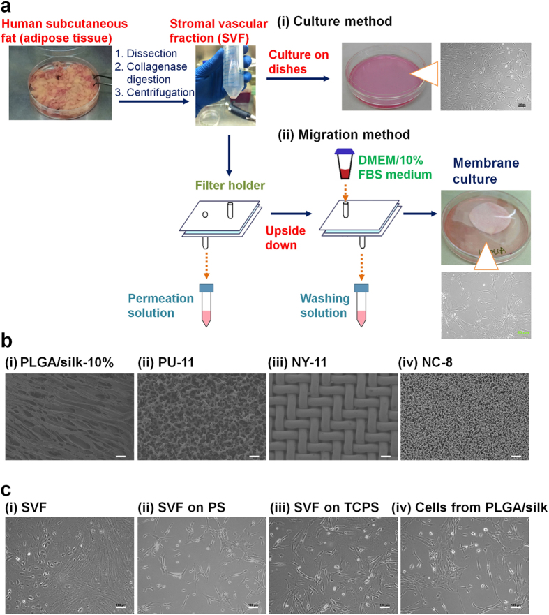
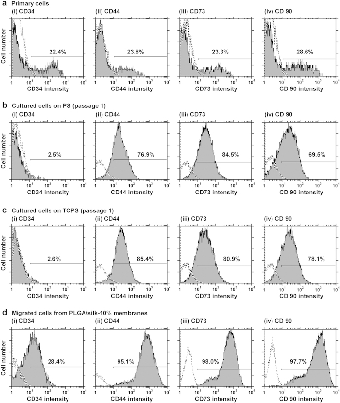
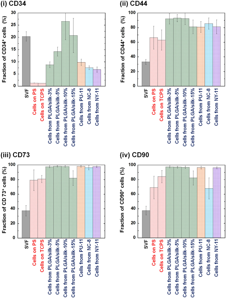
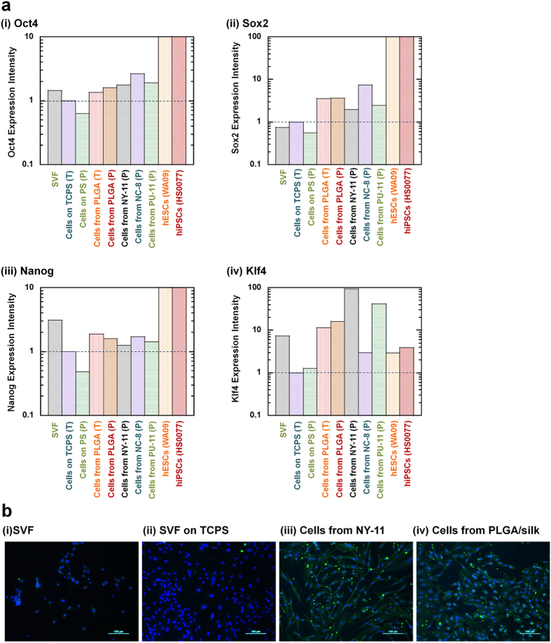
Human adipose-derived stem cells (hADSCs) exhibit heterogeneous characteristics, indicating various genotypes and differentiation abilities. The isolated hADSCs can possess different purity levels and divergent properties depending on the purification methods used. We developed a hybrid-membrane migration method that purifies hADSCs from a fat tissue solution with extremely high purity and pluripotency. A primary fat-tissue solution was permeated through the porous membranes with a pore size from 8 to 25 μm, and the membranes were incubated in cell culture medium for 15-18 days. The hADSCs that migrated from the membranes contained an extremely high percentage (e.g., >98%) of cells positive for mesenchymal stem cell markers and showed almost one order of magnitude higher expression of some pluripotency genes (Oct4, Sox2, Klf4 and Nanog) compared with cells isolated using the conventional culture method.


Figures

Figure 1
Figure 1. Purification of hADSCs from adipose tissue using the hybrid-membrane migration method.
(a) Procedure of the hybrid-membrane migration method. The primary adipose tissue cell solution (SVF) was filtered through the membranes. Subsequently, a washing solution identical to the culture medium was passed through the membranes. The membranes were removed from the membrane holder and inserted into dishes containing culture medium. hADSCs migrated out from the membranes and were expanded in the dishes during culture. (b) Morphology of PLGA/silk-10% (i), polyurethane (PU-11) (ii), nylon mesh filter (NY-11) (iii) and nitrocellulose (NC-8) (iv) membranes used for the hybrid-membrane migration method. The scale bars indicate 100 μm (i) and 40 μm (ii-iv). (c) The morphology of the primary adipose tissue cells (SVF) (i), SVF cells cultured on untreated PS dishes (ii) and on TCPS dishes (iii) for 8 days, and the cells that migrated from PLGA/silk-10% membranes and were subsequently cultured for 15 days after SVF was permeated through the membranes (iv). The scale bars indicate 100 μm.
Figure 2
Figure 2. Purity of hADSCs isolated using the hybrid-membrane migration method.
As analyzed by flow cytometry, the expression of CD34 (i) and MSC markers (CD44 [ii], CD73 [iii], and CD90 [iv]) on the primary adipose tissue cells (SVF) (a), first-passage SVF cells grown in untreated PS (b) and TCPS (c) dishes, and the cells that migrated out from PLGA/silk-10% membranes that were subsequently cultured for 15 days after SVF was permeated through the membranes (d). The dotted lines indicate cells labeled with the isotype controls.
Figure 3
Figure 3. MSC marker expression of hADSCs isolated using the hybrid-membrane migration method.
As analyzed by flow cytometry, the expression of CD34 (i), CD44 (ii), CD73 (iii) and CD90 (iv) on SVF cells, first-passage cells cultured on untreated PS and TCPS, and the cells that migrated out from PLGA/silk, PU-11, NC-8 and NY-11 membranes and were subsequently cultured on untreated PS dishes for 15 days after SVF was permeated through the membranes.
Figure 4
Figure 4. Pluripotency of hADSCs isolated using the hybrid-membrane migration method.
(a) Relative gene expression levels of Oct4 (i), Sox2 (ii), Nanog (iii) and Klf4 (iv) as analyzed by qRT-PCR in the primary adipose tissue cells (SVF), the primary adipose tissue cells (SVF) cultured on untreated PS and TCPS dishes for 8 days (passage one), the cells that migrated out from PLGA/silk-10%, PU-11, NC-8 and NY-11 membranes and were subsequently cultured on untreated PS (P) and TCPS (T) dishes after SVF was permeated through the membranes, hESCs (WA09) and hiPSCs (HS0077). The gene expression was standardized to the expression in the primary adipose tissue cells (SVF) cultured on TCPS. (b) The expression of the pluripotency protein Oct4 in the primary adipose tissue cells (SVF) cultured on untreated PS dishes (i), the primary adipose tissue cells (SVF) cultured on TCPS dishes for 8 days (passage one) (ii), and the cells that migrated out from NY-11 (iii) and PLGA/silk-10% (iv) membranes and were subsequently cultured on untreated PS dishes after SVF was permeated through the membranes, as analyzed by immunostaining for Oct4. Oct4 expression is shown in green, and nuclear staining by Hoechst is shown in blue. The scale bars indicate 100 μm.

References

    1. Thomson J. A. et al. Embryonic stem cell lines derived from human blastocysts. Science 282, 1145–1147 (1998). - PubMed
    1. Takahashi K. et al. Induction of pluripotent stem cells from adult human fibroblasts by defined factors. Cell 131, 861–872 (2007). - PubMed
    1. Higuchi A., Ling Q. D., Ko Y. A., Chang Y. & Umezawa A. Biomaterials for the feeder-free culture of human embryonic stem cells and induced pluripotent stem cells. Chem. Rev. 111, 3021–3034 (2011). - PubMed
    1. Yu J. et al. Induced pluripotent stem cell lines derived from human somatic cells. Science 318, 1917–1920 (2007). - PubMed
    1. Kim J. B. et al. Direct reprogramming of human neural stem cells by OCT4. Nature 461, 649–643 (2009). - PubMed

Publication types