. 2015 May 13;10(5):e0124150.
doi: 10.1371/journal.pone.0124150. eCollection 2015.

A Drosophila model identifies a critical role for zinc in mineralization for kidney stone disease

Affiliations

A Drosophila model identifies a critical role for zinc in mineralization for kidney stone disease

Thomas Chi et al. PLoS One. .

Abstract

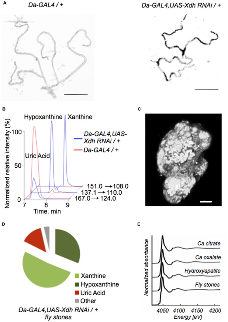
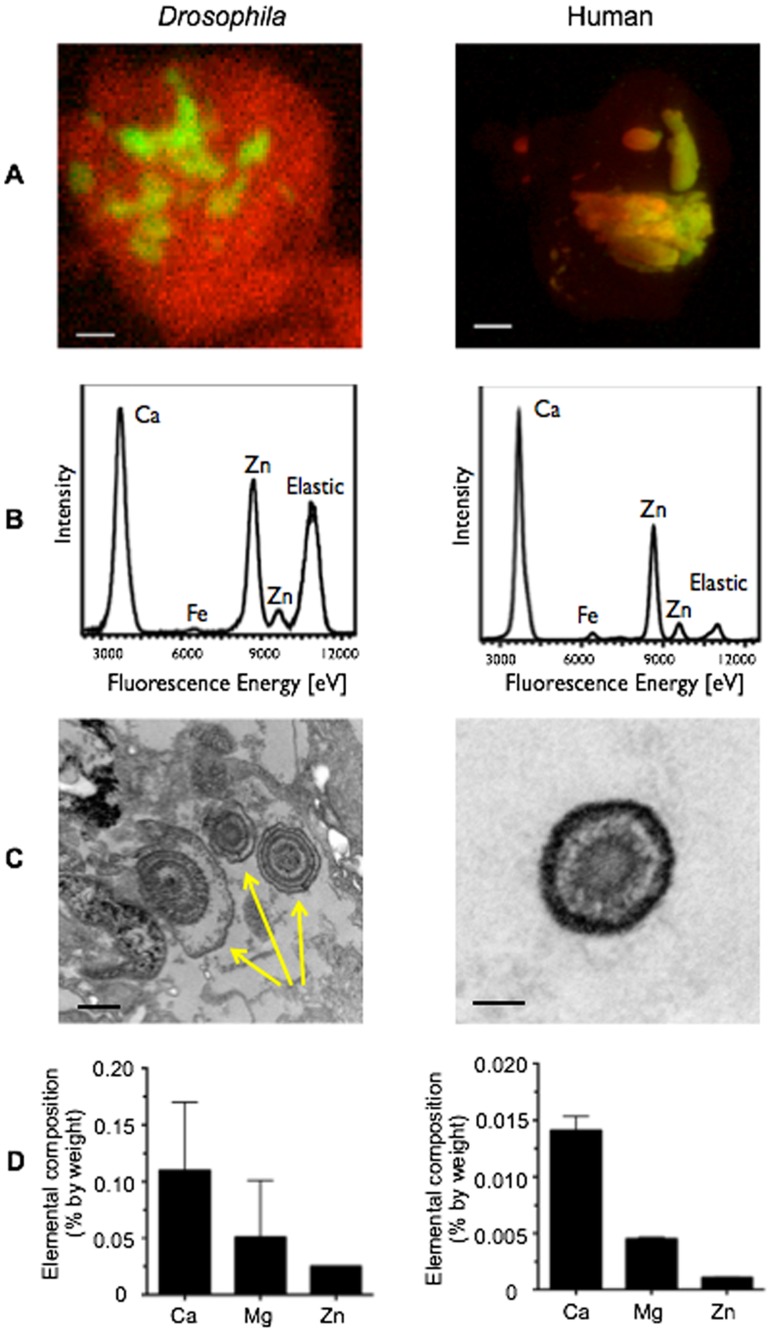
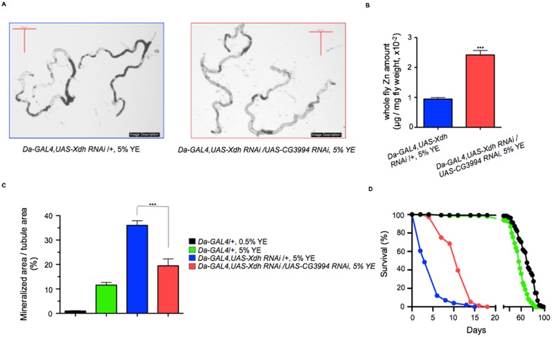
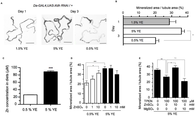
Ectopic calcification is a driving force for a variety of diseases, including kidney stones and atherosclerosis, but initiating factors remain largely unknown. Given its importance in seemingly divergent disease processes, identifying fundamental principal actors for ectopic calcification may have broad translational significance. Here we establish a Drosophila melanogaster model for ectopic calcification by inhibiting xanthine dehydrogenase whose deficiency leads to kidney stones in humans and dogs. Micro X-ray absorption near edge spectroscopy (μXANES) synchrotron analyses revealed high enrichment of zinc in the Drosophila equivalent of kidney stones, which was also observed in human kidney stones and Randall's plaques (early calcifications seen in human kidneys thought to be the precursor for renal stones). To further test the role of zinc in driving mineralization, we inhibited zinc transporter genes in the ZnT family and observed suppression of Drosophila stone formation. Taken together, genetic, dietary, and pharmacologic interventions to lower zinc confirm a critical role for zinc in driving the process of heterogeneous nucleation that eventually leads to stone formation. Our findings open a novel perspective on the etiology of urinary stones and related diseases, which may lead to the identification of new preventive and therapeutic approaches.


Conflict of interest statement

Competing Interests: The authors have declared that no competing interests exist.

Figures

Fig 1
Fig 1. Inhibition of xanthine dehydrogenase leads to ectopic calcification in the fly Malpighian tubules.
(A) Representative tubule images taken from control flies (Da-GAL4/+, left panel) and flies upon Xdh knockdown (Da-GAL4, UAS-Xdh RNAi /+, right panel). Concretions are dark and intraluminal. Scale bars: 500 μm. (B) HPLC-MS (multiple reaction monitoring) analysis of whole flies demonstrates significant reduction in uric acid levels in Da-GAL4, UAS-Xdh RNAi /+ flies compared to control flies with concomitant increase in xanthine and hypoxanthine levels. (C) Confocal microscopy of a fly tubule concretion isolated after Xdh knockdown. The gross appearance resembles that of a miniaturized kidney stone. Scale bar: 20 μm. (D) HPLC-MS (multiple reaction monitoring) analysis of concretions taken from these Da-GAL4, UAS-Xdh RNAi /+ flies demonstrates that they primarily contain xanthine and hypoxanthine with smaller amounts of uric acid and other purine metabolites also present. (E) μXANES demonstrates the presence of hydroxyapatite as the primary calcium salt in Drosophila concretion samples.
Fig 2
Fig 2. Drosophila concretions, human Randall’s plaques, and human kidney stones show enrichment of zinc.
(A) Micro-X-ray fluorescence (μXRF) maps of Da-GAL4, UAS-Xdh RNAi /+ concretions (left panel, scale bar: 10 μm) and human Randall’s plaques (right panel, scale bar: 100 μm) demonstrate the presence of zinc in red and calcium in green. (B) μXRF elemental analysis of the samples from (A) demonstrate similar elemental composition for both Da-GAL4, UAS-Xdh RNAi /+ concretions (left panel) and human Randall’s plaques (right panel), including the presence of calcium (Ca), iron (Fe), and zinc (Zn). (C) Transmission electron microscopy imaging of concretions in the lumen of the Malpighian tubule demonstrates the presence of ring-like structures, as indicated by the yellow arrows (left panel, scale bar: 500 μm). Ring structures with homologous appearance are seen in Randall’s plaques taken from a human renal papilla biopsy material (right panel, scale bar: 100 μm) when imaged in a similar fashion. (D) ICP-OES analysis of pooled fly concretion samples from 300 dissected tubule specimens demonstrates the presence of calcium (Ca), magnesium (Mg), and zinc (Zn) (left panel, n = 2 biological replicates). These levels are mirrored in human xanthine stone samples (right panel, n = 2 biological replicates). Data shown are the mean ± SEM.
Fig 3
Fig 3. Inhibition of zinc transporters inhibits concretion formation in a fly model for concretion formation.
(A) Gross photomicrographs of Malpighian tubule concretions isolated from Da-GAL4, UAS-Xdh RNAi /+ flies fed on a 5% YE diet (left panel) demonstrate significant reduction in concretion formation after simultaneous inhibition of the zinc transporter CG3994 (right panel). Scale bars: 500 μm. (B) Upon simultaneous RNAi inhibition of xanthine dehydrogenase and CG3994, male flies were fed 5% YE. After 2 days, flies were anesthetized with CO2, their body weights measured and whole flies homogenized and analyzed with ICP-OES. Whole-fly levels of Zn were significantly higher after zinc transport inhibition (red bar) compared to flies expressing Xdh inhibition alone (blue bar, ***p<0.001, Student’s t-test, n = 3). Data shown are the mean ± SEM. (C) Measuring the area of the tubule lumen occupied by mineralized concretions, Da-GAL4 /+ control flies produce significantly more concretions after feeding 5% YE compared to 0.5% YE (green versus black bars). With silencing of Xdh, tubule concretion production increases dramatically (blue bar), but simultaneous inhibition of xanthine dehydrogenase and CG3994 resulted in significant reduction in concretion accumulation compared to Da-GAL4, UAS-Xdh RNAi /+ flies (red bar) (***p <0.001, one way ANOVA with Bonferroni post-hoc test, n = 14–43). (D) Similarly, while Da-GAL4, UAS-Xdh RNAi /+ flies exhibit significantly reduced survivorship (blue line), simultaneous inhibition of CG3994 with xanthine dehydrogenase rescued survivorship when compared with Da-GAL4, UAS-Xdh RNAi /+ animals on 5% YE (red line, logrank test, n = 76–97).
Fig 4
Fig 4. Zinc modulates stone formation in a fly model for ectopic calcification.
(A) Typical tubule images taken from Da-GAL4, UAS-Xdh-RNAi /+ flies at eclosion (Day 1, 1.5% YE) and after feeding 2 days on high yeast (Day 3, 5% YE) and low yeast (Day 3, 0.5% YE) diets. (B) The extent of tubular mineralization is quantified as the percentage of the tubule lumen occupied by mineralized material (*p <0.001, Student’s t-test, n = 14–48). Scale bars: 500 μm. Data shown are the mean ± SEM. (C) The amount of Zn in the components of a standard Drosophila diet (corn meal, yeast, sugar and agar) was measured with ICP-OES for both 0.5% YE and 5% YE diets. 0.5% YE is comprised of corn meal 8.5 g, yeast 0.5 g, sugar 5 g, and agar 0.46 g in 100 ml deionized distilled water. 5% YE is composed of the same components as 0.5% YE with the exception of 5 g yeast added instead of 0.5 g yeast (***p<0.001, student t-test, n = 3). (D) Da-GAL4, UAS-Xdh RNAi / + flies fed on 0.5% YE and 5% YE diets were supplemented with different doses of Zn. This led to a dose-dependent increase in accumulation of concretions on 0.5% YE feeding (*p <0.05, ***p <0.001, one way ANOVA with Bonferroni post-hoc test, n = 10–48). Zn supplementation had no effect on altering concretion formation in 5% YE-fed animals, which may reflect a saturation of the calcification process under these conditions. (E) Supplementation of 5% YE diet with the zinc chelator TPEN reduced concretion formation. This effect could be reversed with the addition of 10 mM Zn but not 10 mM Mg (*p <0.05, **p <0.01, one way ANOVA with Bonferroni post-hoc test, n = 10–43). Data shown are the mean ± SEM.

References

    1. Yuan Q, Sato T, Densmore M, Saito H, Schüler C, Erben RG, et al. (2012) Deletion of PTH rescues skeletal abnormalities and high osteopontin levels in Klotho-/- mice. PLoS Genet 8: e1002726 10.1371/journal.pgen.1002726 - DOI - PMC - PubMed
    1. Nikdin H, Olsson M-L, Hultenby K, Sugars RV (2012) Osteoadherin accumulates in the predentin towards the mineralization front in the developing tooth. PLoS ONE 7: e31525 10.1371/journal.pone.0031525 - DOI - PMC - PubMed
    1. Giachelli CM (1999) Ectopic calcification: gathering hard facts about soft tissue mineralization. Am J Pathol 154: 671–675. 10.1016/S0002-9440(10)65313-8 - DOI - PMC - PubMed
    1. Evan AP, Coe FL, Lingeman JE, Worcester E (2005) Insights on the pathology of kidney stone formation. Urol Res 33: 383–389. 10.1007/s00240-005-0488-0 - DOI - PubMed
    1. Duer MJ, Friscić T, Proudfoot D, Reid DG, Schoppet M, Shanahan CM, et al. (2008) Mineral surface in calcified plaque is like that of bone: further evidence for regulated mineralization. Arterioscler Thromb Vasc Biol 28: 2030–2034. 10.1161/ATVBAHA.108.172387 - DOI - PubMed

Publication types

MeSH terms