Review
. 2015 May 11;5(2):848-64.
doi: 10.3390/biom5020848.

Structural biology of bacterial RNA polymerase

Affiliations
Review

Structural biology of bacterial RNA polymerase

Katsuhiko S Murakami. Biomolecules. .

Abstract

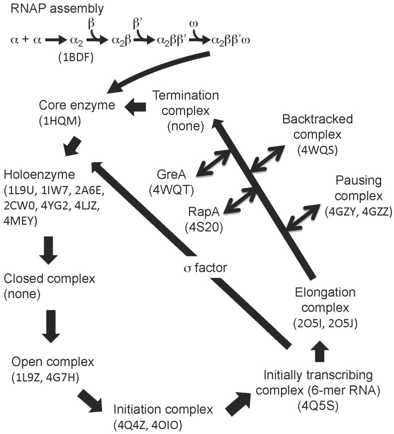
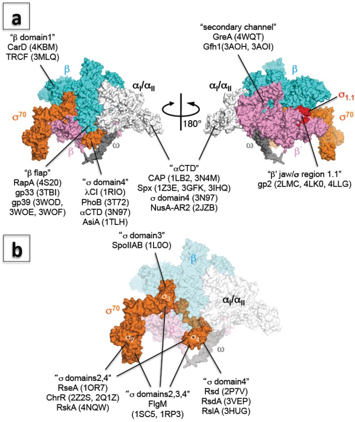
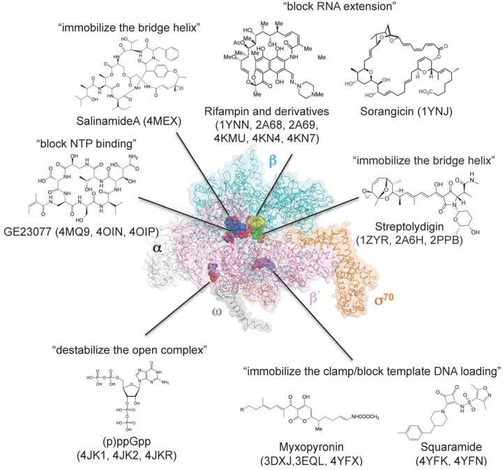
Since its discovery and characterization in the early 1960s (Hurwitz, J. The discovery of RNA polymerase. J. Biol. Chem. 2005, 280, 42477-42485), an enormous amount of biochemical, biophysical and genetic data has been collected on bacterial RNA polymerase (RNAP). In the late 1990s, structural information pertaining to bacterial RNAP has emerged that provided unprecedented insights into the function and mechanism of RNA transcription. In this review, I list all structures related to bacterial RNAP (as determined by X-ray crystallography and NMR methods available from the Protein Data Bank), describe their contributions to bacterial transcription research and discuss the role that small molecules play in inhibiting bacterial RNA transcription.

Keywords: NMR; X-ray crystallography; anti-σ factor; bacterial RNA polymerase; core enzyme; holoenzyme; inhibitor antibiotic; transcription; transcription factor; σ factor.


Figures

Figure 1
Figure 1
RNAP assembly and transcription cycle. The figure shows the general scheme for bacterial RNAP assembly and the transcription cycle. PDB codes of RNAP structures (in brackets) belonging to each phase of the transcription cycle.
Figure 2
Figure 2
(a) Three-dimensional representation of the interaction between RNAP and transcription factors. The E. coli RNAP holoenzyme is shown as a molecular surface representation (α subunits: white; β subunit: cyan; β’ subunit: pink; ω subunit: gray; σ70: orange; σ region 1.1: red). Transcription factors binding sites are indicated in double quotation marks and PDB codes of structures are shown in brackets; (b) Three-dimensional representation of the interaction between σ and anti-σ factors. E. coli RNAP holoenzyme is shown as a molecular surface representation, and only the core enzyme is partially transparent (α subunits: white; β subunit: cyan; β’ subunit: pink; ω subunit: gray; σ70: orange). Targets of anti-σ factors are indicated in double quotation marks and PDB codes of structures are shown in brackets.
Figure 3
Figure 3
Three-dimensional representation of targets of small-molecule RNAP inhibitors. E. coli RNAP holoenzyme is depicted as an α-carbon backbone traced with partially transparent molecular surfaces (α subunits: white; β subunit: cyan; β' subunit: pink; ω subunit: gray; σ70: orange). Small-molecule inhibitors bound to RNAP are depicted as CPK models. Chemical structures of small-molecule inhibitors and mechanisms of transcription inhibition are indicated.

References

    1. Murakami K.S., Darst S.A. Bacterial RNA polymerases: The wholo story. Curr. Opin. Struct. Biol. 2003;13:31–39. doi: 10.1016/S0959-440X(02)00005-2. - DOI - PubMed
    1. Jun S.H., Warner B.A., Murakami K.S. RNA polymerase reaction in bacteria. In: Lane W.J.L.D., editor. Encyclopedia of Biological Chemistry. Academic Press; Waltham, MA, USA: 2013. pp. 167–172.
    1. Fuchs E., Zillig W., Hofschneider P.H., Preuss A. Preparation and properties of RNA-polymerase particles. J. Mol. Biol. 1964;10:546–550. doi: 10.1016/S0022-2836(64)80076-0. - DOI - PubMed
    1. Tsuji S., Suzuki K., Imahori K. Crystallisation of DNA-dependent RNA polymerase from Thermus thermophilus HB 8. Nature. 1976;261:725–726. doi: 10.1038/261725a0. - DOI - PubMed
    1. Jeon Y.H., Negishi T., Shirakawa M., Yamazaki T., Fujita N., Ishihama A., Kyogoku Y. Solution structure of the activator contact domain of the RNA polymerase alpha subunit. Science. 1995;270:1495–1497. doi: 10.1126/science.270.5241.1495. - DOI - PubMed

Publication types

MeSH terms

LinkOut - more resources