Review
. 2015 Aug;1854(8):1054-70.
doi: 10.1016/j.bbapap.2015.05.001. Epub 2015 May 10.

Breaking a pathogen's iron will: Inhibiting siderophore production as an antimicrobial strategy

Affiliations
Review

Breaking a pathogen's iron will: Inhibiting siderophore production as an antimicrobial strategy

Audrey L Lamb. Biochim Biophys Acta. 2015 Aug.

Abstract

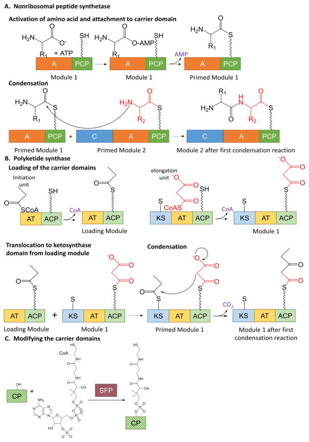
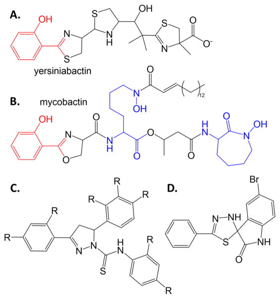
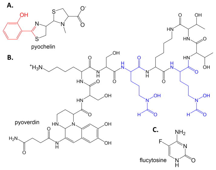
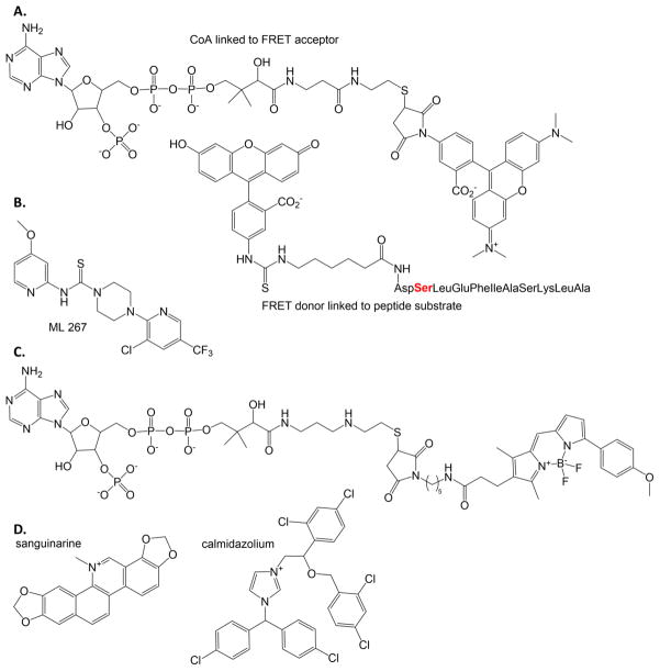
The rise of antibiotic resistance is a growing public health crisis. Novel antimicrobials are sought, preferably developing nontraditional chemical scaffolds that do not inhibit standard targets such as cell wall synthesis or the ribosome. Iron scavenging has been proposed as a viable target, because bacterial and fungal pathogens must overcome the nutritional immunity of the host to be virulent. This review highlights the recent work toward exploiting the biosynthetic enzymes of siderophore production for the design of next generation antimicrobials.

Keywords: NRPS-independent synthetase; Nonribosomal peptide synthetase; Polyketide synthase; Siderophore.


Figures

Figure 1
Figure 1. Siderophore biosynthesis
A. Nonribosomal peptide synthetase chain elongation. The adenylation domain activates an amino acid and attaches it to the carrier domain, thereby priming the module (top line). The condensation domain forms a peptide bond between two primed modules (bottom line). The process continues with more modules in an assembly line fashion. A = adenylation, C = condensation, PCP = peptidyl carrier protein. The wavy line in the PCP domain denotes the phosphopantethienyl post-translational modification. B. Polyketide synthase bond formation. The acyl transferase domains load acyl groups onto the carrier domains of the loading module and module 1 (top). The acyl group from the loading module is transferred to the ketosynthase domain of module 1 thereby priming the module (bottom left). The ketosynthase domain performs the condensation reaction (bottom right). The growing chain is transferred to the ketosynthase domain of the next module for the assembly line to continue. AT = acyl transferase, ACP = acyl carrier protein, KS = ketosynthase. The wavy line in the ACP domain denotes the phosphopantethienyl post-translational modification. The short, straight line in the KS domain denotes an active site cysteine. C. Post translational modification of carrier domains to generate phosphopantetheinyl swinging arm. CP = carrier protein is striped in shades of green to represent that this is common to both NRPS (dark green) and PKS (light green) carrier domains. SFP is a promiscuous PPTase from Bacillus subtilis commonly used to perform this reaction in vitro.
Figure 2
Figure 2. Siderophore mimics
Salicylate-capped siderophores yersiniabactin (A) and mycobactin (B). The salicylate caps are shown as red. Hydroxylysine residues are blue. C. Scaffold for siderophore mimics with antimicrobial activity against Y. pestis and M. tuberculosis [20]. D. Spiro-indoline-thiadiazole inhibitor that converts to a merocyanine metal chelator and has antimicrobial activity against E. coli [21].
Figure 3
Figure 3. Repurposing drugs
The P. aeruginosa siderophores pyochelin (A) and pyoverdin (B). In pyochelin, the salicylate cap is again red. In pyoverdin, the formyl-hydroxyornithine residues are blue. C. Flucytosine.
Figure 4
Figure 4. Phosphopantetheinyl transferase probes and inhibitor
A. The FRET probes developed to determine inhibitors of PPTase enzymes. [28] The serine where the PPTase attaches the FRET acceptor to the FRET donor-labeled peptide is shown in red. B. ML267 inhibitor of Sfp-PPTase identified by high throughput screening using the FRET assay using probes in part A. [31, 32] C. BODIPY-TMR fluorescence polarization probe developed to assay PPTase inhibitors. [33] D. The two most inhibitory compounds of the PPTase from M. tuberculosis are shown, determined when using the fluorescence polarization assay where the probe is the FRET acceptor in part A. [34, 35]
Figure 5
Figure 5. Inhibitors of salicylate and dihydroxybenzoate adenylation enzymes
A. Salicyl-AMS, a rationally designed reaction intermediate analogue. [36]. B. Benzoyl-AMN [43]. C. DHB-hydroxamoyl adenylate [44]. D. Inhibitor from high throughput screen. [46]. E. Three structures of BasE are overlayed: BasE with DHB-AMS bound is the orange cartoon with green stick inhibitor (PDB ID: 3O82), with a triazole derivative of DHB-AMS shown in cyan sticks (PDB ID: 3O83), and with inhibitor from part D shown in magenta sticks (PDB ID: 3O84). F. The area in the box in part E is shown in stereo (all stereo images are wall-eye or divergent stereo). Note the unexpected binding mode of the magenta HTS inhibitor relative the green and cyan substrate analogue inhibitors. Structure figures made in PyMOL [121].
Figure 6
Figure 6. Dihydroxbenzoate-capped siderophores
Bacillobactin from B. subtilis, acinetobactin from A. baumannii, enterobactin from E. coli, and vibrobactin from V. cholera. DHB caps are shown in pink.
Figure 7
Figure 7. Inhibitors of adenylation domains in NRPS modules
A. Macrocyclic inhibitor of cysteine adenylation domain in HWMP2 for yersiniabactin biosynthesis. [50] B. Mechanism based inhibitor used to trap adenylation and carrier domains in stable conformation. The one shown here was designed for the DHB-specific EntE [53]. C. The structure of the carrier domain (green cartoon) and adenylation domain (orange) with the vinylsulfonamide inhibitor (magenta sticks) of PA1221 from P. aeruginosa, a two domain NRPS specific for the incorporation of valine (PDB ID: 4DG9). D. Close up of the active site showing the covalent linkage of the adenylation domain inhibitor (similar to that in part B) to the phosphopantetheinyl post translation modification of the carrier domain.
Figure 8
Figure 8. NRPS-independent siderophore (NIS) synthetase inhibitor
A. Condensation of citric acid (blue) with diaminopropionic acid (red) by NIS synthetase SbnE from S. aureus for the production of staphyloferrin. B. Condensation of citric acid (blue) with spermidine (green) by NIS synthetase AsbA from B. anthrasis for the production of petrobactin. C. Micromolar antibiotic inhibitor of SbnE and AsbA [61].
Figure 9
Figure 9. Isochorismate synthase, salicylate synthase, isochorismate-pyruvate lyase
A. Salicylate production for the generation of salicylate-capped siderophores. B. Potential transition state analogue inhibitor for isochorismate synthase activity. [68] C. Salicylate synthase inhibitor from HTS. [82] D. Isochorismate-pyruvate lyase inhibitor from HTS that also is effective against salicylate synthase and chorismate mutase. [86]. E. The structure of the salicylate synthase from Y. enterocolitica (Irp9, purple cartoon, PDB ID: 2FN1) with the catalytic Mg ion (green sphere) and products salicylate and pyruvate (purples sticks) was used to align two inhibitor-bound structures of the salicylate synthase from M. tuberculosis, MbtI. An MbtI structure with the required Mg has never been determined. The structure of MbtI with the aromatized isochorismate analogue known as AMT is shown in yellow sticks (PDB ID: 3ST6). MbtI with methyl-AMT is shown in cyan sticks (PDB ID: 3VEH). The methyl group addition is at the –ene of the pyruvylenol tail. Cartoons for 3ST6 and 3VEH are not included for the sake clarity. F. Close up of the active site is shown in stereo illustrating the binding modes of the inhibitors relative to products. Note that AMT (yellow) binds similarly to the substrate/products, with the salicyl ring analogue coordinated to the Mg2+. By contrast, additions to the pyruvylenol tail cause a flipping of the binding mode (methyl-AMT as one example, cyan) with the salicyl ring analogue now binding in the location where the pyruvate product is normally found.
Figure 10
Figure 10. Ornithine hydroxylase
A. Ferrichrome and ferricrocin siderophores from A. fumigatus. The formyl-hydroxyornithine residues are blue. B. ADP-TAMRA chromophore designed for a high throughput assay for the flavin-dependent N-hydroxylating monooxygenases, such as ornithine and lysine hydroxylases.
Figure 11
Figure 11. Inhibiting ornithine decarboxylase to generate novel siderophores
A. Putrebactin, the natural siderophore of S. putrefaciens with putrescine (diaminobutane, red) incorporated. B. When ornithine decarboxylase is inhibited, preventing the production of putrescine, then S. putrefaciens makes desferrioxamine, using cadaverine (diaminopentane, green) instead. C. When ornithine decarboxylase is inhibited and an external source of diaminobutene (blue) is added to the culture, then an unsaturated form of putrebactin is formed.
Figure 12
Figure 12. Reporter substrates and inhibitors for PvdQ
A. Substrate analogue probes. 4-nitrophenyl myristate provides an absorbance readout, whereas 4-methyl-umbelliferyl laurate is fluorescent when cleaved. [103] B. Inhibitor of PvdQ identified by high throughput screening and SAR. [106, 107] C. Rationally designed transition state analogue inhibitor [108]. D. The PvdQ structures with ML318 (PDB ID: 4K2G, inhibitor in green sticks) and tridecylboronic acid (PDB ID: 4M1J, inhibitor cyan) are overlayed. Only the latter cartoon is shown for clarity. E. Stereoview of active site with inhibitors bound. Note that the boronic acid inhibitor (boron is pink) forms a covalent transition state analogue with the serine nucleophile (grey sticks).

Similar articles

Cited by

References

    1. The White House Executive Order -- Combating Antibiotic-Resistant Bacteria. https://www.whitehouse.gov/the-press-office/2014/09/18/executive-order-c....
    1. Centers for Disease Control and Prevention Antibiotic Resistance Solutions Initiative. http://www.cdc.gov/drugresistance/solutions-initiative/index.html.
    1. World Health Organization. Antimicrobial Resistance: Global Report on Surveillance. 2014.
    1. Clatworthy AE, Pierson E, Hung DT. Targeting virulence: a new paradigm for antimicrobial therapy. Nat Chem Biol. 2007;3(9):541–8. - PubMed
    1. Skaar EP. The battle for iron between bacterial pathogens and their vertebrate hosts. PLoS pathogens. 2010;6(8):e1000949. - PMC - PubMed

Publication types

MeSH terms