. 2015 Aug 21;6(24):20525-39.
doi: 10.18632/oncotarget.3890.

Type II transglutaminase stimulates epidermal cancer stem cell epithelial-mesenchymal transition

Affiliations

Type II transglutaminase stimulates epidermal cancer stem cell epithelial-mesenchymal transition

Matthew L Fisher et al. Oncotarget. .

Abstract

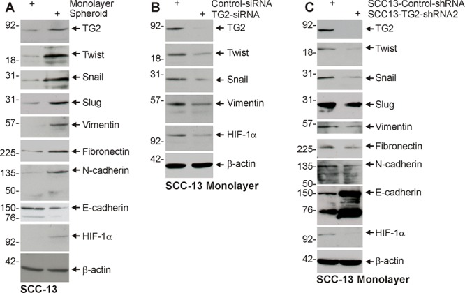
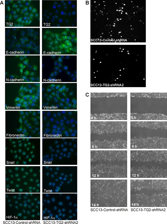
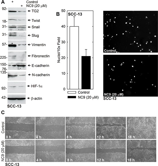
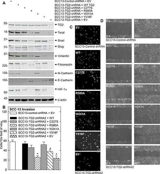
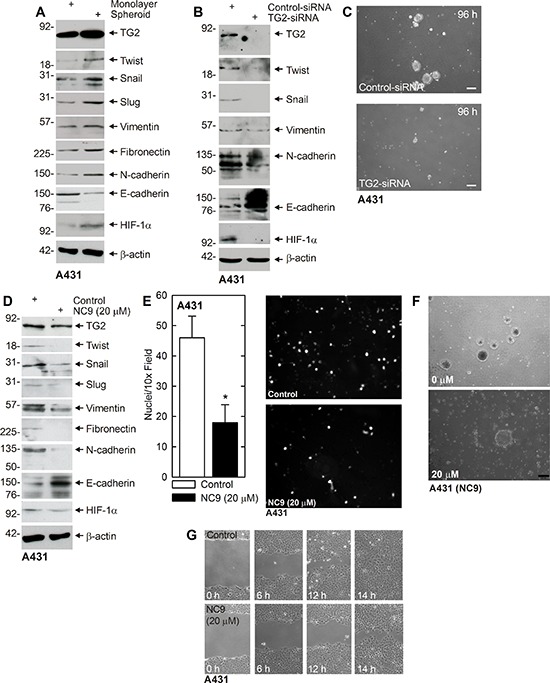
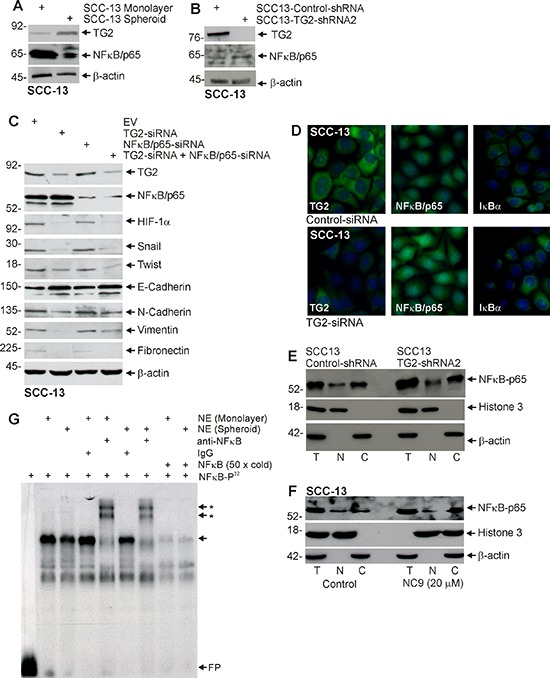
Type II transglutaminase (TG2) is a multifunctional protein that has recently been implicated as having a role in ECS cell survival. In the present study we investigate the role of TG2 in regulating epithelial mesenchymal transition (EMT) in ECS cells. Our studies show that TG2 knockdown or treatment with TG2 inhibitor, results in a reduced EMT marker expression, and reduced cell migration and invasion. TG2 has several activities, but the most prominent are its transamidase and GTP binding activity. Analysis of a series of TG2 mutants reveals that TG2 GTP binding activity, but not the transamidase activity, is required for expression of EMT markers (Twist, Snail, Slug, vimentin, fibronectin, N-cadherin and HIF-1α), and increased ECS cell invasion and migration. This coupled with reduced expression of E-cadherin. Additional studies indicate that NFϰB signaling, which has been implicated as mediating TG2 impact on EMT in breast cancer cells, is not involved in TG2 regulation of EMT in skin cancer. These studies suggest that TG2 is required for maintenance of ECS cell EMT, invasion and migration, and suggests that inhibiting TG2 GTP binding/G-protein related activity may reduce skin cancer tumor survival.

Keywords: TG2; cancer stem cell; epidermal squamous cell carcinoma; epithelial mesenchymal transition; type II transglutaminase.


Figures

Figure 1
Figure 1. TG2 and EMT marker expression is enriched in ECS cells, and TG2 is required for EMT
A. ECS cells have elevated TG2 and mesenchymal markers. SCC-13 cells (40,000 per well) were grown in spheroid medium in attached (monolayer) and non-attached (spheroid, ECS cells). Cells were harvested after 10 d and lysates were prepared for electrophoresis and detection of the indicated epitopes. B. SCC-13 cells were electroporated with control- or TG2-siRNA and cultured in spheroid medium as monolayer cultures. After 72 h extracts were prepared to assay TG2 and EMT markers. C. SCC13-Control-shRNA and SCC13-TG2-shRNA2 cells were grown in spheroid media for 10 d, harvested and lysates prepared for immune-detection of TG2 and EMT markers. Similar results were observed in three separate experiments.
Figure 2
Figure 2. TG2 is required for EMT marker expression and invasion/migration
A. SCC13-Control-shRNA and SCC13-TG2-shRNA2 cells were grown in spheroid medium as spheroids in non-attached culture for 8 d, and then plated as monolayers in 12 well plates (100,000 cells/well). After overnight attachment, the cells were fixed, permeabilized and incubated with antibodies specific for the indicated epitopes. B. SCC13-Control-shRNA and SCC13-TG2-shRNA2 cells were seeded on the upper chamber atop a matrigel-coated membrane in 1 ml of growth medium in a Millicell chamber. After migration, the membrane was removed, rinsed, fixed and DAPI stained. Nuclei were counted on the underside of the membrane using an inverted fluorescent microscope. C. SCC13-Control-shRNA and SCC13-TG2-shRNA2 cells (2 million) were plated on 100 mm dishes in spheroid medium in monolayer conditions. The confluent monolayers were scratched with a 10 μl pipette tip to create a wound and the released cells were removed. Images were taken at 0–14 h after the initial scratch. Similar results were observed in three experiments.
Figure 3
Figure 3. NC9 reduces EMT protein expression and invasion/migration
A. SCC-13 cells (500,000) were plated in monolayer culture in spheroid medium in 100 mm dishes. Fresh spheroid medium containing 0 or 20 μM NC9 was added at time zero and again at 24 h. After 48 h lysates were collected for electrophoresis and immunoblot. B. SCC-13 cells were pretreated with 0 or 20 μM NC9 for 2 h and then harvested and plated (25,000 cells) in the upper chamber in 1 ml of medium, above a matrigel-coated membrane, in a Millicell chamber. After 30 h, the inside of membrane was harvested and the nuclei of migrated cells on the bottom of the membrane were counted using an inverted fluorescent microscope. The values are mean ± SEM, n = 3 (p, 0.05). C. SCC-13 (2 million) cells were plated in spheroid medium in 100 mm dishes and grown as monolayer cultures. Confluent monolayers were then scratched with a 10 μl pipette tip and wound width was monitored for 0–18 h. Similar results were observed in each of three experiments.
Figure 4
Figure 4. TG2 GTP binding/G-protein function is necessary for EMT
A. TG2 impact on EMT marker expression. SSC13-Control-shRNA or SCC13-TG2-shRNA2 cells were electroporated with 3 μg of empty vector (EV) or vector encoding the indicated TG2 proteins. The cells were then grown as monolayer cultures in spheroid medium for three days. The cells were then harvested and extracts prepared for electrophoresis and immunoblot detection of the indicated epitopes. Similar results were observed in three experiments. B. SCC13-TG2-shRNA2 cells were electroporated with plasmids encoding wild-type TG2, mutant TG2, or empty vector and grown in spheroid medium as monolayers. At 48 h post-electroporation, SCC13-Control-shRNA and SCC13-TG2-shRNA2 cells were harvested and seeded in the upper chamber above a matrigel-coated membrane in 1 ml of spheroid medium containing 1% serum in a Millicell chamber. The lower chamber included spheroid medium containing 10% serum. Nuclei of migrated cells were stained and counted on the lower surface of the membrane using an inverted fluorescent microscope. The values are mean ± SEM, n = 3 (p < 0.01). C. Images of DAPI-stained membrane. D. SCC13-Control-shRNA and SCC13-TG2-shRNA2 cells were electroporated with empty vector (EV) or vector encoding the indicated TG2 proteins and grown in spheroid medium as monolayers. After 24 h, 2 million cells were plated in spheroid media on 100 mm dishes in monolayer conditions. The confluent monolayer cultures were then scratched with a 10 μl pipette tip and width of the wound was monitored at 0–18 h to assess closure. Similar results were observed in each of three experiments.
Figure 5
Figure 5. TG2, EMT markers and EMT response in A431 cells
A. A431-derived ECS cells have elevated TG2 and mesenchymal markers. A431 cells (40,000 cells/well) were grown in spheroid media in monolayer conditions or as non-attached spheroids (ECS cells) for 10 d. Cells were harvested and lysates prepared for immunoblot. B. A431 cells were electroporated with 3 μg of control- or TG2-siRNA and at 72 h extracts were prepared to assay TG2 and EMT marker level. C. A431-derived ECS cells were electroporated with 3 μg of the indicated siRNA and then grown as spheroids for 96 h. Similar results were observed in three separate experiments. D. A431 cells (500,000 cells/well) were plated in monolayer culture in spheroid media in 100 mm dishes. The following day, at time zero, 0 or 20 μM NC9 was added with fresh addition of medim and NC9 at 24 h. At 48 h the cells were harvested and extracts prepared for immunoblot. E. A431 cells were pretreated in 0 or 20 μM NC9 for 2 h and then harvested and 25,000 cells were seeded in the upper chamber above a matrigel-coated membrane in 1 ml of growth medium containing 1% FCS in a Millicell chamber. The lower chamber was filled with growth medium containing 10% FCS. After 30 h, the membrane was removed, washed and stained with DAPI, and an inverted fluorescent microscope was used to count the nuclei of migrated cells. F. A431 (40,000) cells were plated in spheroid media in non-attached conditions. On d 8, spheroids were treated with 0 or 20 μM NC9 and images were taken after 48 h to monitor spheroid fragmentation. G. A431 (2 million) cells were plated in spheroid media on 100 mm dishes and grown as monolayers. Confluent monolayers were then scratched with a 10 μl pipette tip, rinsed, and width of the wound was monitored for 0–14 h. Similar results were observed in each of three experiments.
Figure 6
Figure 6. NFκB expression is not required for EMT
A. ECS cells have reduced NFκB-p65 levels. SCC-13 cells (40,000) were grown in spheroid medium in attached (monolayer) and non-attached (spheroid, ECS cells) conditions. Lysates were prepared after 10 d in culture for electrophoresis and detection of indicated epitopes. B. SCC13-Control-shRNA and SCC13-TG2-shRNA2 cells were grown in spheroid medium as monolayers. After 10 d in culture, lysates were collected for detection of TG2 and NFκB-p65. C. SCC-13 cells were electroporated with control-, NFκB-p65- or TG2-siRNA and then cultured as monolayers in spheroid medium. After 72 h extracts were prepared to assay TG2, NFκB-p65 and EMT markers. D. SCC-13 cells were electroporated with empty vector Control- or TG2-siRNA and plated in monolayer culture. The following day cells were fixed, permeabilized and incubated with antibodies specific for the indicated epitopes. Similar results were observed in each of three experiments. E. An equal number of cell equivalents of total (T), nuclear (N), and cytosolic (C) extract, prepared from SCC13-Control-shRNA and SCC13-TG2-shRNA2 (TG2 knockdown) cells, was electrophoresed for immunoblot detection of NFκB, histone 3 (nuclear marker) and β-actin (cytoplasmic marker). Similar results were observed in three separate experiments. F. Total (T), nuclear (N), and cytosolic (C) extract was prepared from 8 day spheroids (ECS cells) treated with 0 or 20 μM NC9 for 48 h. An equal number of cell equivalents of total, nuclear and cytosol extract was electrophoresed for immunoblot detection with the indicated antibodies. Similar results were observed in three separate experiments. G. Nuclear extract (NE), prepared from non-stem cancer cells (monolayer) or ECS cells (spheroids), was incubated with 32P-NFκB probe and electrophoresed on a non-denaturing 6% acrylamide gel. Various groups were incubated with IgG, anti-NFkB or a 50-fold (50× cold) molar excess of radioinert NFkB probe prior to electrophoresis. FP indicates free 32P-NFκB probe, the arrow indicates the gel mobility shift and the arrows with asterisks indicate the anti-dependent supershifted bands.
Figure 7
Figure 7. NFκB is not required for ECS cell migration and invasion
A, B. NFκB-knockdown does not suppress ECS cell matrigel invasion. SCC-13 cells were treated with Control-, TG2-, NFκB- or a combination of siRNA and then seeded in the upper chamber above a matrigel-coated membrane in 1 ml of growth medium containing 1% FCS in a Millicell chamber. The lower chamber was filled with growth medium containing 10% FCS. After 30 h, the membrane was removed, washed and stained with DAPI, and an inverted fluorescent microscope was used to count the nuclei of migrated cells. C. NFκB-knockdown does not impede ECS cell migration. SCC-13 cells (2 million) cells were plated in spheroid media on 100 mm dishes and grown as monolayers. Confluent monolayers were then scratched with a 10 μl pipette tip, rinsed, and width of the wound was monitored for 0–18 h. Similar results were observed in each of three experiments.

Similar articles

Cited by

References

    1. Christenson LJ, Borrowman TA, Vachon CM, Tollefson MM, Otley CC, Weaver AL, Roenigk RK. Incidence of basal cell and squamous cell carcinomas in a population younger than 40 years. JAMA. 2005;294:681–690. - PubMed
    1. Moller R, Reymann F, Hou-Jensen K. Metastases in dermatological patients with squamous cell carcinoma. Arch Dermatol. 1979;115:703–705. - PubMed
    1. Adhikary G, Grun D, Kerr C, Balasubramanian S, Rorke EA, Vemuri M, Boucher S, Bickenbach JR, Hornyak T, Xu W, Fisher ML, Eckert RL. Identification of a population of epidermal squamous cell carcinoma cells with enhanced potential for tumor formation. PLoS One. 2013;8:e84324. - PMC - PubMed
    1. Samatov TR, Tonevitsky AG, Schumacher U. Epithelial-mesenchymal transition: focus on metastatic cascade, alternative splicing, non-coding RNAs and modulating compounds. Mol Cancer. 2013;12:107. - PMC - PubMed
    1. Chaffer CL, Thompson EW, Williams ED. Mesenchymal to epithelial transition in development and disease. Cells Tissues Organs. 2007;185:7–19. - PubMed

Publication types

MeSH terms

Substances