

Spontaneous electrical activities at myofascial trigger points at different stages of recovery from injury in a rat model
- PMID: 25971282
- PMCID: PMC4552908
- DOI: 10.1136/acupmed-2014-010666
Spontaneous electrical activities at myofascial trigger points at different stages of recovery from injury in a rat model
Erratum in
-
Correction.Acupunct Med. 2015 Oct;33(5):434. doi: 10.1136/acupmed-2014-010666corr1. Acupunct Med. 2015. PMID: 26447110 Free PMC article. No abstract available.
Abstract
Background: Spontaneous electrical activity (SEA) is a feature of myofascial trigger points (MTrPs), which can either be latent or active. However, SEA at different stages of recovery from MTrPs remains unclear.
Objective: To investigate the temporal changes in the nature of SEA after generation of MTrPs in a rat model.
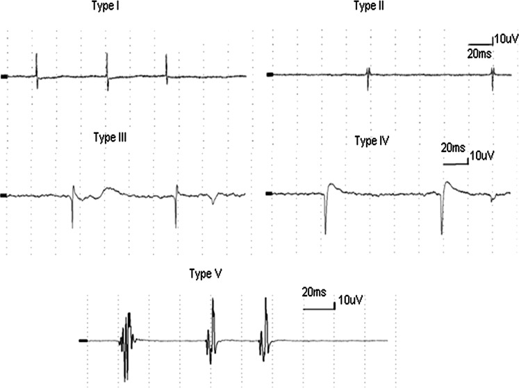
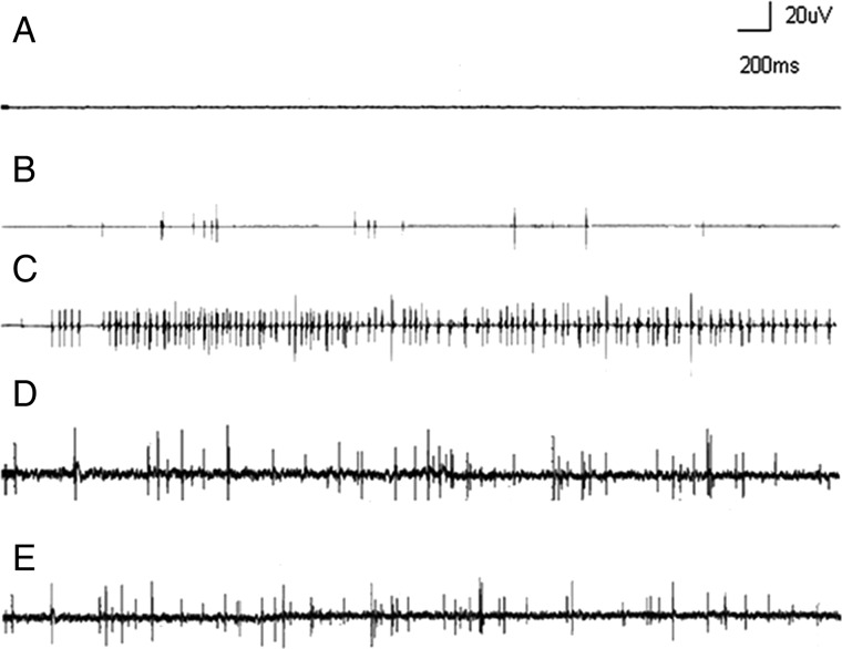
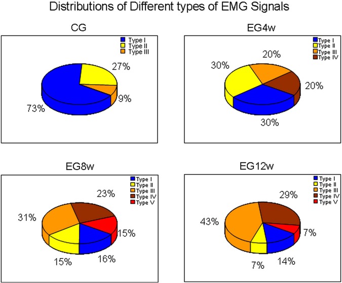
Methods: 32 rats were divided into four groups: 24 rats were assigned to experimental groups (EGs), which underwent the MTrP modelling intervention and 8 were allocated to a control group (CG). All EG rats received a blunt strike to the left vastus medialis combined with eccentric exercise for 8 weeks. After modelling, the EG rats were subdivided into three groups with total recovery times of 4, 8 and 12 weeks (EG-4w, EG-8w and EG-12w, respectively). Taut bands (TBs) with and without the presence of active MTrPs were identified in the left hind limb muscles of all rats, verified by SEA and further examined with electromyography recordings. Myoelectrical signals were also categorised into one of five types.
Results: CG rats had fewer TBs than EG rats and EGs showed variable frequencies of SEA. SEA frequencies were higher in EG-4w than in EG-8w and EG-12w groups (240.57±72.9 vs 168.14±64.5 and 151.63±65.4, respectively, p<0.05) and were significantly greater in all EGs than in the CG (55.75±21.9). Relative to CG rats, amplitudes and durations of electrical potentials in the EG were only increased in the EG-8w and EG-12w groups. Types IV and V myoelectrical signals were never seen in latent MTrPs and type V signals did not occur in EG-4w rats.
Conclusions: Increasing recovery periods following a MTrP modelling intervention in rats are characterised by different frequencies and amplitudes of SEA from TBs.
Trial registration number: 2014012.
Keywords: MYOFASCIAL PAIN; PAIN RESEARCH.
Published by the BMJ Publishing Group Limited. For permission to use (where not already granted under a licence) please go to http://group.bmj.com/group/rights-licensing/permissions.
Figures
References
-
- Simons DG, Travell JG, Simons LS. Myofascial pain and dysfunction: the trigger point manual.Vol.1. Upper half of the body. Baltimore, MD: Lippincott Williams & Wilkins, 1999.
-
- Dommerholt J, Bron C, Franssen J. Myofascial trigger points: an evidence-informed review. J Man Manip Ther 2006;14:203–21. 10.1179/106698106790819991 - DOI
-
- Dommerholt J. Dry needling in orthopedic physical therapy practice. Orthop Phys Ther Pract 2004;16:15–20.
Publication types
MeSH terms
LinkOut - more resources
Full Text Sources
Other Literature Sources
