. 2015 May 12;107(8):djv123.
doi: 10.1093/jnci/djv123. Print 2015 Aug.

Targeting Focal Adhesion Kinase and Resistance to mTOR Inhibition in Pancreatic Neuroendocrine Tumors

Affiliations

Targeting Focal Adhesion Kinase and Resistance to mTOR Inhibition in Pancreatic Neuroendocrine Tumors

Rony A François et al. J Natl Cancer Inst. .

Abstract

Background: Focal adhesion kinase (FAK) mediates survival of normal pancreatic islets through activation of AKT. Upon malignant transformation of islet cells into pancreatic neuroendocrine tumors (PanNETs), AKT is frequently overexpressed and mutations in the AKT/mTOR pathway are detected. Because mTOR inhibitors rarely induce PanNET tumor regression, partly because of feedback activation of AKT, novel combination strategies are needed to target FAK/AKT/mTOR signaling.

Methods: We characterized the activation of FAK in PanNETs using immunohistochemistry and Western blot analysis and tested the FAK inhibitor PF-04554878 in human PanNET cells in vitro and in vivo (at least three mice per group). In addition, we evaluated the effect of combined FAK and mTOR inhibition on PanNET viability and apoptosis. All statistical tests were two-sided.

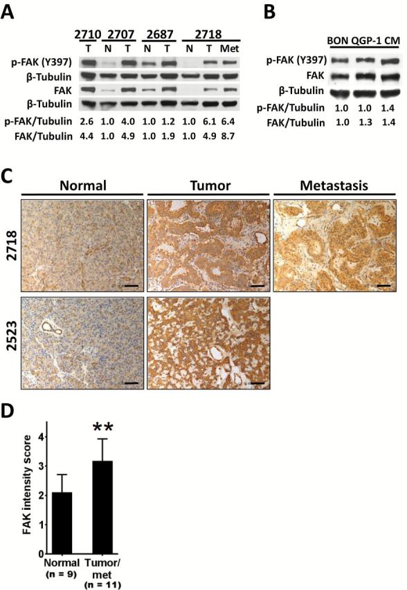
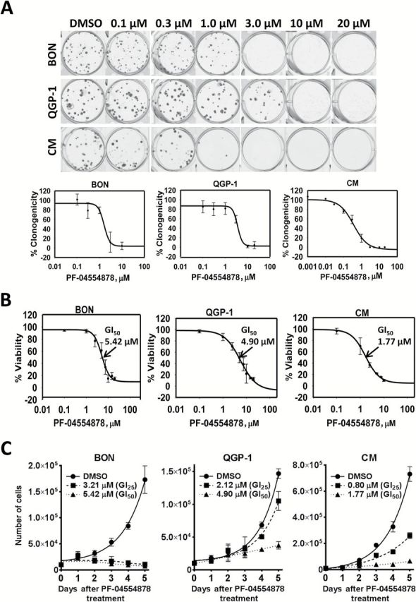
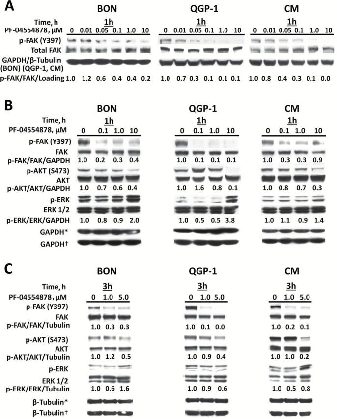
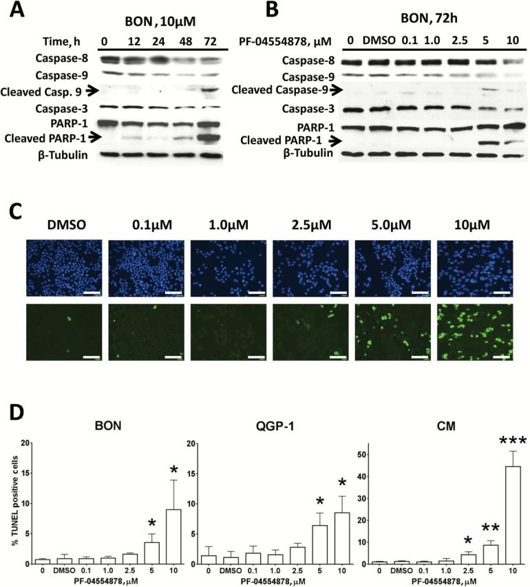
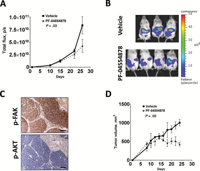
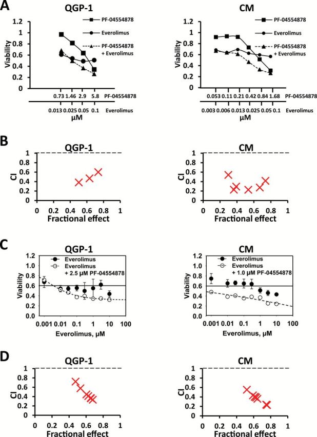
Results: We found that FAK is overexpressed and hyperphosphorylated in human PanNETs and that PF-04554878 strongly inhibited FAK (Tyr397) autophosphorylation in a dose-dependent manner. We found that PF-04554878 inhibited cell proliferation and clonogenicity and induced apoptosis in PanNET cells. Moreover, oral administration of PF-04554878 statistically significantly reduced tumor growth in a patient-derived xenograft model of PanNET (P = .02) and in a human PanNET xenograft model of peritoneal carcinomatosis (P = .03). Importantly, PF-04554878 synergized with the mTOR inhibitor everolimus by preventing feedback AKT activation.

Conclusions: We demonstrate for the first time that FAK is overexpressed in PanNETs and that inhibition of FAK activity induces apoptosis and inhibits PanNET proliferation. We found that the novel FAK inhibitor PF-04554878 synergizes with everolimus, a US Food and Drug Administration-approved agent for PanNETs. Our findings warrant the clinical investigation of combined FAK and mTOR inhibition in PanNETs.


Figures

Figure 1.
Figure 1.
Focal adhesion kinase (FAK) expression in pancreatic neuroendocrine tumors (PanNETs). A) Immunoblot analysis of Y397 phospho- and total FAK in PanNET patient tumors (T) and/or matched metastatic sample (Met) as compared with matched normal pancreas (N). Numbers (2710, etc.) indicate samples obtained from the same patient. For densitometric analysis, results were normalized to tubulin and are expressed as fold induction over matched normal sample. For the unmatched tumor sample, results are expressed as fold induction over the mean of the other three normal samples. B) Immunoblot analysis of phospho- (Y397) and total FAK in human PanNET cell lines. Tubulin is shown as a loading control, and densitometric analyses are expressed as fold induction over BON cells. C) Representative images of FAK immunohistochemistry staining in human PanNET samples (scale bar = 200 µm). D) Analysis of FAK immunohistochemistry staining intensity in nine normal samples and 11 matched PanNET tumor samples (including one unmatched tumor and one matched metastasis sample) as determined by a masked pathologist using a two-tailed Student’s t test (**P < .01). Error bars represent standard deviation of the mean. FAK = focal adhesion kinase.
Figure 2.
Figure 2.
Effect of PF-04554878, a novel inhibitor of focal adhesion kinase (FAK) kinase activity, on growth of human pancreatic neuroendocrine tumor (PanNET) cells. A) Dose-response effect of PF-04554878 on colony formation of BON, QGP-1, and CM cells at low cell density after two- to four-week treatment as determined by the clonogenicity assay, and quantification of colonies for BON, QGP-1, and CM cells as determined by mechanical tally counter. Percent clonogenicity was calculated by normalizing colony counts to DMSO control. Error bars represent standard deviation of the mean. B) Viability of BON, QGP-1, and CM cells treated for 72 hours with increasing doses of PF-04554878 as determined by MTT assay. Percent viability was calculated by normalizing absorbance to DMSO control. Error bars represent standard deviation of the mean. C) Proliferation growth curves of BON, QGP-1, and CM cells treated for five days with DMSO control or PF-04554878 at GI25 or GI50 concentrations. Error bars represent standard deviation of the mean. GI50 = 50% growth inhibition, GI25 = 25% growth inhibition.
Figure 3.
Figure 3.
Effect of PF-04554878 on phosphorylation of focal adhesion kinase (FAK), AKT, and ERK. A) Immunoblot analysis of phosphorylated (Y397) and total FAK protein levels after one hour of treatment with increasing doses of PF-04554878 in human pancreatic neuroendocrine tumor cell lines. B) Effect of one hour or (C) three hours of PF-04554878 treatment on FAK, AKT, and ERK1/2 activation as determined by immunoblot analysis of phosphorylated FAK (Y397), AKT (S473), and ERK1/2 (Thr202/204) in comparison with total protein levels. Tubulin or GAPDH were used as loading controls. *Loading control for phosphoproteins. †Loading control for total proteins. FAK = focal adhesion kinase.
Figure 4.
Figure 4.
Effect of PF-04554878 on apoptosis in human pancreatic neuroendocrine tumor (PanNET) cells. A) Time course immunoblot analysis of the effects of 10 µM of PF-04554878 on induction of apoptosis as determined by caspase 3, 8, and 9 levels, as well as PARP-1 cleavage. B) Effect of increasing doses of PF-04554878 on caspases 3, 8, and 9 activation and PARP-1 cleavage in BON cells after 72 hours of treatment. C) Representative images of TUNEL staining for apoptosis in three independent dose-response experiments in CM cells following 72 hours of treatment with PF-04554878 (scale bar = 200 µm). D) Graphical quantification of PF-04554878 induction of apoptosis in human PanNET cell lines as determined by TUNEL staining. For each experiment, five images of each slide were analyzed, and the percentage of apoptotic cells was calculated by counting the number of TUNEL positive nuclei (green) in relation to total nuclei stained with DAPI (blue). Results are composite of three independent experiments. A two-tailed Student’s t test was used to compare each group with DMSO control (*P < .05, **P < .01, ***P < .001). Error bars represent standard deviation of the mean.
Figure 5.
Figure 5.
Effect of PF-04554878 on growth of human human pancreatic neuroendocrine tumors (PanNETs) in vivo. A) Effect of daily oral administration of PF-04554878 on growth of CM-Luc cells in NOD/SCID mice as measured by bioluminescent luciferase signal intensity in CM-Luciferase model of PanNET carcinomatosis. Fold induction bioluminescent signal intensity was calculated for each mouse by normalizing bioluminescent signal value to first measurement (Day 10 post-injection). Based on bioluminescent imaging, mice were assigned on Day 15 to vehicle control (n = 3) or 50mg/kg of PF-04554878 (n = 4). B) Bioluminescent imaging of mice on final day of study (Day 26). C) Immunohistochemical staining of FAK (Y397) and AKT (S473) phosphorylation in PanNET patient-derived xenograft tumor model (scale bar = 200 µm). D) Effect of daily oral administration of PF-04554878 on growth of human PanNET patient-derived xenograft transplant model in NSG mice. Animals were implanted with a 3 x 3 x 3mm piece of a human PanNET tumor sample 10 days prior to treatment assignment and commencement (Day 1) with vehicle control or 50mg/kg of PF-04554878. Tumors were measured when palpable using calipers, and tumor volume was calculated using the formula V = L2 x W x π/6. Statistical analysis of the effect of treatment intervention as compared with control was performed using a repeated measures two-tailed two-way analysis of variance. Error bars represent standard deviation of the mean.
Figure 6.
Figure 6.
Effect of dual targeting of focal adhesion kinase (FAK) and mTOR on pancreatic neuroendocrine tumor (PanNET) cell growth. A) Effect of PF-0455878 and everolimus alone and in combination using the fixed molar ratio method in QGP1 and CM cells. PF-0455878 and everolimus were combined in a constant EC50 ratio, followed by serial one-half dilutions of this combination. B) CalcuSyn graph of combination index values (CI) and fraction of cells affected (Fa) from fixed molar ratio combination of PF-04554878 and everolimus in QGP-1 and CM cells. C) Effect of everolimus alone and in combination with a single dose of PF-04554878 using the varying molar ratio method in QGP1 and CM cells. A single nonapoptogenic dose of PF-04554878 was combined with increasing concentrations of everolimus. D) CalcuSyn graph of combination index values (CI) and fraction of cells affected (Fa) from varying molar ratio combination of PF-04554878 and everolimus in QGP-1 and CM cells.

Similar articles

Cited by

References

    1. Zhang J, Francois R, Iyer R, Seshadri M, Zajac-Kaye M, Hochwald SN. Current understanding of the molecular biology of pancreatic neuroendocrine tumors. J Natl Cancer Inst. 2013;105(14):1005–1017. - PMC - PubMed
    1. Jiao Y, Shi C, Edil BH, et al. DAXX/ATRX, MEN1, and mTOR pathway genes are frequently altered in pancreatic neuroendocrine tumors. Science. 2011;331(6021):1199–1203. - PMC - PubMed
    1. Oberstein PE, Saif MW. Safety and efficacy of everolimus in adult patients with neuroendocrine tumors. Clin Med Insights Oncol. 2012;6:41–51. - PMC - PubMed
    1. Yao JC, Shah MH, Ito T, et al. Everolimus for advanced pancreatic neuroendocrine tumors. N Engl J Med. 2011;364(6):514–523. - PMC - PubMed
    1. Sankhala K, Giles FJ. Potential of mTOR inhibitors as therapeutic agents in hematological malignancies. Expert Rev Hematol. 2009;2(4):399–414. - PubMed

Publication types

MeSH terms