

Expression of folate receptors alpha and beta in normal and cancerous gynecologic tissues: correlation of expression of the beta isoform with macrophage markers
- PMID: 25971554
- PMCID: PMC4464638
- DOI: 10.1186/s13048-015-0156-0
Expression of folate receptors alpha and beta in normal and cancerous gynecologic tissues: correlation of expression of the beta isoform with macrophage markers
Abstract
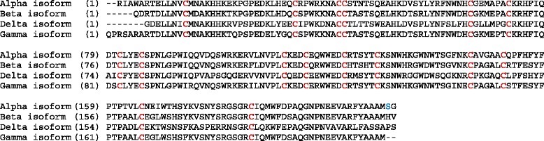
Background: Folate receptor alpha (FOLR1/FRA) is expressed in a number of epithelial cancers and in particular epithelial ovarian cancer (EOC), especially of the serous histotype. Recent studies have shown that EOC originates from the fallopian tube fimbriae rather than from epithelial cells lining the ovary. We have previously shown by immunohistochemistry a strong correlation between FRA expression in EOC and normal and fallopian adenocarcinoma. Folate receptor beta (FOLR2/FRB) has been described to be expressed by macrophages both in inflammatory disorders and certain epithelial cancers. Given the high sequence identity of these two folate receptor family members we sought to investigate the architectural and cell-specific expression of these two receptors in gynecologic tissues.
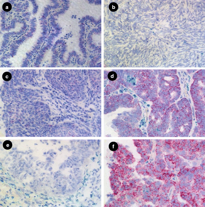
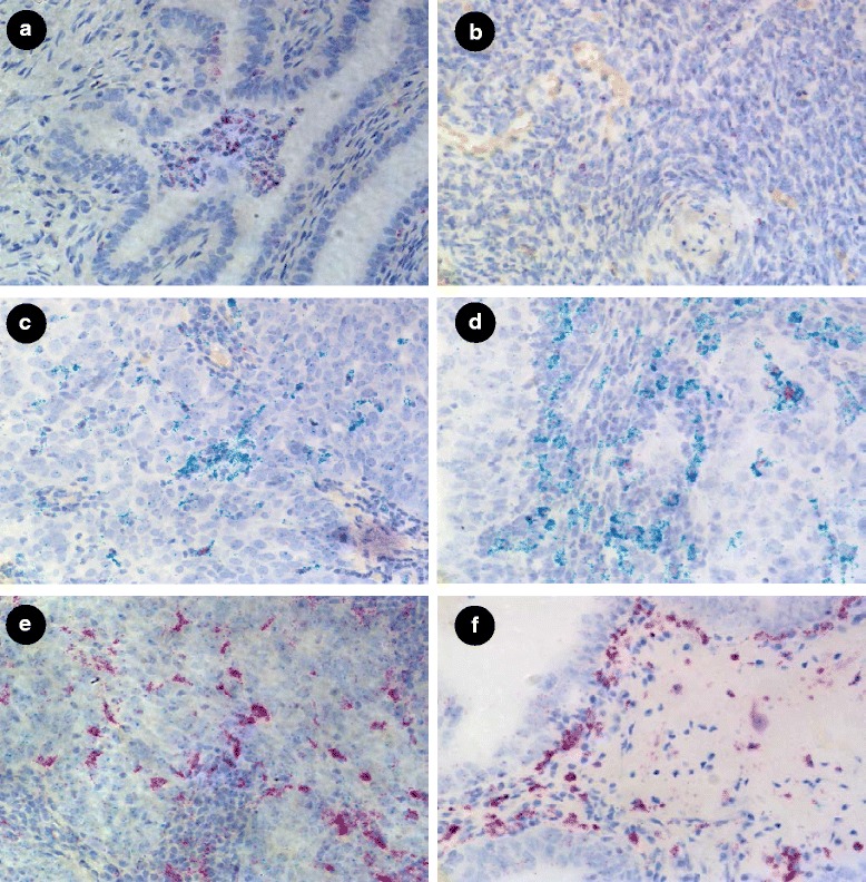
Methods: RNA scope, a novel chromogenic in situ hybridization assay tool, was used to examine expression of the alpha (FOLR1) and beta (FOLR2) isoforms of folate receptor relative to each other as well as to the macrophage markers CD11b and CD68, in samples of normal fallopian tube and fallopian adenocarcinoma as well as normal ovary and EOC.
Results: We demonstrated expression of both FOLR1 and FOLR2 in EOC, normal fallopian tube and fallopian adenocarcinoma tissue while very little expression of either marker was observed in normal ovary. Furthermore, FOLR2 was shown to be expressed almost exclusively in macrophages, of both the M1 and M2 lineages, as determined by co-expression of CD11b and/or CD68, with little or no expression in epithelial cells.
Conclusions: These findings further substantiate the hypothesis that the cell of origin of EOC is tubal epithelium and that the beta isoform of folate receptor is primarily restricted to macrophages. Further, macrophages expressing FOLR2 may represent tumor associated or infiltrating macrophages (TAMs) in epithelial cancers.
Figures
Similar articles
-
Gene expression analyses support fallopian tube epithelium as the cell of origin of epithelial ovarian cancer.Int J Mol Sci. 2013 Jul 1;14(7):13687-703. doi: 10.3390/ijms140713687. Int J Mol Sci. 2013. PMID: 23880844 Free PMC article.
-
[Significance and expression of PAX8, PAX2, p53 and RAS in ovary and fallopian tubes to origin of ovarian high grade serous carcinoma].Zhonghua Fu Chan Ke Za Zhi. 2017 Oct 25;52(10):687-696. doi: 10.3760/cma.j.issn.0529-567X.2017.10.008. Zhonghua Fu Chan Ke Za Zhi. 2017. PMID: 29060967 Chinese.
-
Epithelialization of mouse ovarian tumor cells originating in the fallopian tube stroma.Oncotarget. 2016 Oct 4;7(40):66077-66086. doi: 10.18632/oncotarget.11808. Oncotarget. 2016. PMID: 27602775 Free PMC article.
-
Role of Fallopian Tubes in the Development of Ovarian Cancer.J Minim Invasive Gynecol. 2017 Feb;24(2):230-234. doi: 10.1016/j.jmig.2016.12.007. Epub 2016 Dec 19. J Minim Invasive Gynecol. 2017. PMID: 28007588 Review.
-
Ovarian serous carcinogenesis from tubal secretory cells.Histol Histopathol. 2015 Nov;30(11):1295-302. doi: 10.14670/HH-11-645. Epub 2015 Jul 15. Histol Histopathol. 2015. PMID: 26174492 Review.
Cited by
-
Folate receptor-α targeted near-infrared fluorescence imaging in high-risk endometrial cancer patients: a tissue microarray and clinical feasibility study.Oncotarget. 2017 Dec 11;9(1):791-801. doi: 10.18632/oncotarget.23155. eCollection 2018 Jan 2. Oncotarget. 2017. PMID: 29416655 Free PMC article.
-
One-carbon metabolism and nucleotide biosynthesis as attractive targets for anticancer therapy.Oncotarget. 2017 Apr 4;8(14):23955-23977. doi: 10.18632/oncotarget.15053. Oncotarget. 2017. PMID: 28177894 Free PMC article. Review.
-
Niclosamide targets the dynamic progression of macrophages for the resolution of endometriosis in a mouse model.Commun Biol. 2022 Nov 11;5(1):1225. doi: 10.1038/s42003-022-04211-0. Commun Biol. 2022. PMID: 36369244 Free PMC article.
-
Silencing the FOLR2 Gene Inhibits Cell Proliferation and Increases Apoptosis in the NCI-H1650 Non-Small Cell Lung Cancer Cell Line via Inhibition of AKT/Mammalian Target of Rapamycin (mTOR)/Ribosomal Protein S6 Kinase 1 (S6K1) Signaling.Med Sci Monit. 2018 Nov 11;24:8064-8073. doi: 10.12659/MSM.911384. Med Sci Monit. 2018. PMID: 30415267 Free PMC article.
-
The folate receptor β as a macrophage-mediated imaging and therapeutic target in rheumatoid arthritis.Drug Deliv Transl Res. 2019 Feb;9(1):366-378. doi: 10.1007/s13346-018-0589-2. Drug Deliv Transl Res. 2019. PMID: 30280318 Free PMC article. Review.
References
-
- Hough CD, Cho KR, Zonderman AB, Schwartz DR, Morin PJ. Coordinately up-regulated genes in ovarian cancer. Cancer Res. 2001;61:3869–76. - PubMed
MeSH terms
Substances
LinkOut - more resources
Full Text Sources
Other Literature Sources
Medical
Research Materials
