

Affective facilitation of early visual cortex during rapid picture presentation at 6 and 15 Hz
- PMID: 25971598
- PMCID: PMC4666108
- DOI: 10.1093/scan/nsv058
Affective facilitation of early visual cortex during rapid picture presentation at 6 and 15 Hz
Erratum in
-
Corrigendum to: Affective facilitation of early visual cortex during rapid picture presentation at 6 and 15 Hz.Soc Cogn Affect Neurosci. 2017 Jun 1;12(6):1022-1023. doi: 10.1093/scan/nsx024. Soc Cogn Affect Neurosci. 2017. PMID: 28444367 Free PMC article. No abstract available.
Abstract
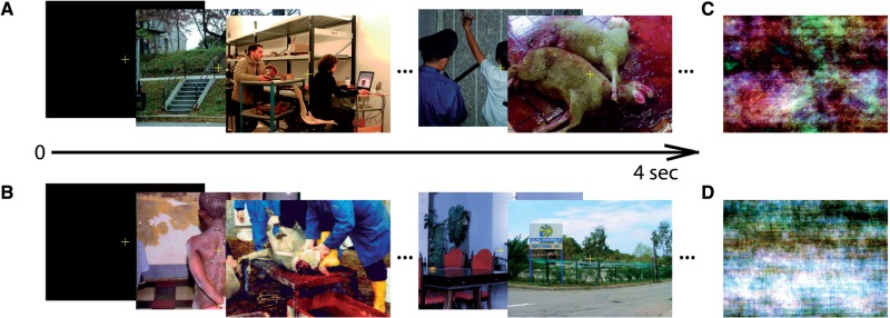
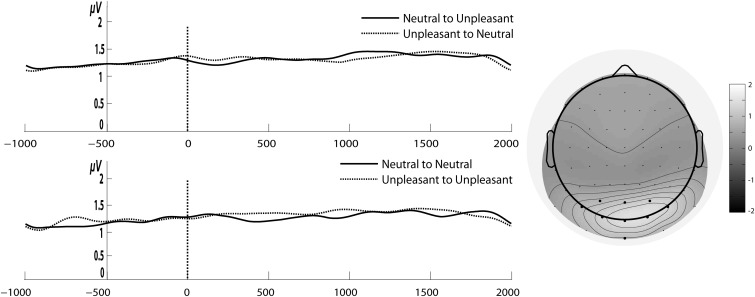
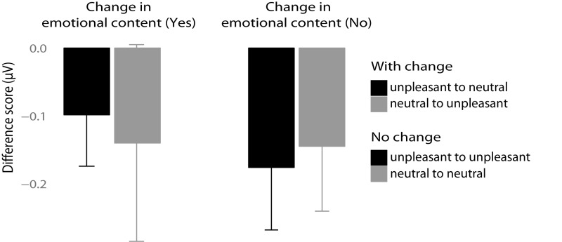
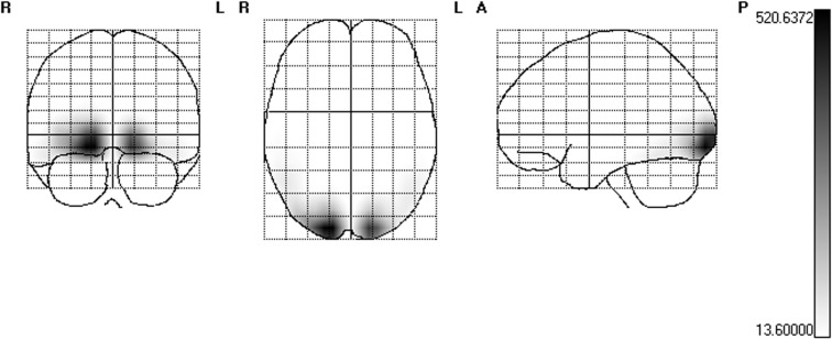
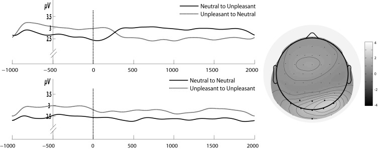
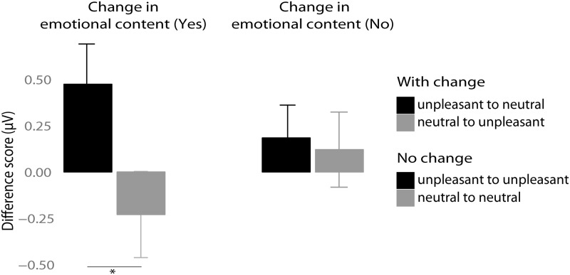
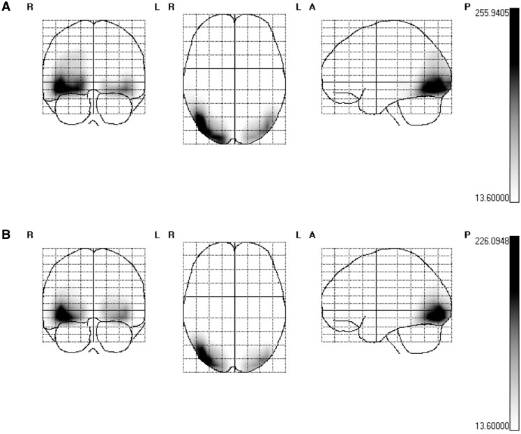
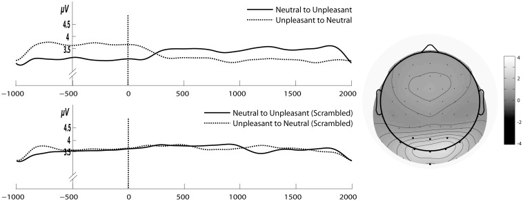
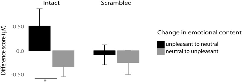
The steady-state visual evoked potential (SSVEP), a neurophysiological marker of attentional resource allocation with its generators in early visual cortex, exhibits enhanced amplitude for emotional compared to neutral complex pictures. Emotional cue extraction for complex images is linked to the N1-EPN complex with a peak latency of ∼140-160 ms. We tested whether neural facilitation in early visual cortex with affective pictures requires emotional cue extraction of individual images, even when a stream of images of the same valence category is presented. Images were shown at either 6 Hz (167 ms, allowing for extraction) or 15 Hz (67 ms per image, causing disruption of processing by the following image). Results showed SSVEP amplitude enhancement for emotional compared to neutral images at a presentation rate of 6 Hz but no differences at 15 Hz. This was not due to featural differences between the two valence categories. Results strongly suggest that individual images need to be displayed for sufficient time allowing for emotional cue extraction to drive affective neural modulation in early visual cortex.
Keywords: SSVEP; attention; emotional cue extraction; human early visual cortex.
© The Author (2015). Published by Oxford University Press. For Permissions, please email: journals.permissions@oup.com.
Figures
Similar articles
-
Attentional bias to affective faces and complex IAPS images in early visual cortex follows emotional cue extraction.Neuroimage. 2015 May 15;112:254-266. doi: 10.1016/j.neuroimage.2015.03.052. Epub 2015 Mar 25. Neuroimage. 2015. PMID: 25818682
-
Slow biasing of processing resources in early visual cortex is preceded by emotional cue extraction in emotion-attention competition.Hum Brain Mapp. 2014 Apr;35(4):1477-90. doi: 10.1002/hbm.22267. Epub 2013 Mar 1. Hum Brain Mapp. 2014. PMID: 23450516 Free PMC article.
-
Competition for attentional resources between low spatial frequency content of emotional images and a foreground task in early visual cortex.Psychophysiology. 2017 Mar;54(3):429-443. doi: 10.1111/psyp.12792. Epub 2016 Dec 19. Psychophysiology. 2017. PMID: 27990660
-
Steady-state visual evoked potentials as a research tool in social affective neuroscience.Psychophysiology. 2016 Dec;53(12):1763-1775. doi: 10.1111/psyp.12768. Epub 2016 Oct 4. Psychophysiology. 2016. PMID: 27699794 Free PMC article. Review.
-
Dynamics of emotional effects on spatial attention in the human visual cortex.Prog Brain Res. 2006;156:67-91. doi: 10.1016/S0079-6123(06)56004-2. Prog Brain Res. 2006. PMID: 17015075 Review.
Cited by
-
Perceptual Difficulty Regulates Attentional Gain Modulations in Human Visual Cortex.J Neurosci. 2023 May 3;43(18):3312-3330. doi: 10.1523/JNEUROSCI.0519-22.2023. Epub 2023 Mar 24. J Neurosci. 2023. PMID: 36963848 Free PMC article.
-
Walking through Architectural Spaces: The Impact of Interior Forms on Human Brain Dynamics.Front Hum Neurosci. 2017 Sep 27;11:477. doi: 10.3389/fnhum.2017.00477. eCollection 2017. Front Hum Neurosci. 2017. PMID: 29033807 Free PMC article.
-
Rapid processing of neutral and angry expressions within ongoing facial stimulus streams: Is it all about isolated facial features?PLoS One. 2020 Apr 24;15(4):e0231982. doi: 10.1371/journal.pone.0231982. eCollection 2020. PLoS One. 2020. PMID: 32330160 Free PMC article.
-
Visual cortex responses reflect temporal structure of continuous quasi-rhythmic sensory stimulation.Neuroimage. 2017 Feb 1;146:58-70. doi: 10.1016/j.neuroimage.2016.11.043. Epub 2016 Nov 17. Neuroimage. 2017. PMID: 27867090 Free PMC article.
-
Bringing color to emotion: The influence of color on attentional bias to briefly presented emotional images.Cogn Affect Behav Neurosci. 2017 Oct;17(5):1028-1047. doi: 10.3758/s13415-017-0530-z. Cogn Affect Behav Neurosci. 2017. PMID: 28699142
References
-
- Alonso-Prieto E., Belle G.V., Liu-Shuang J., Norcia A.M., Rossion B. (2013). The 6 Hz fundamental stimulation frequency rate for individual face discrimination in the right occipito-temporal cortex. Neuropsychologia, 51(13), 2863–75. - PubMed
-
- Andersen S.K., Fuchs S., Müller M.M. (2009). Effects of feature-selective and spatial attention at different stages of visual processing. Journal of Cognitive Neuroscience, 23(1), 238–46. - PubMed
-
- Andersen S.K., Hillyard S.A., Müller M.M. (2008). Attention facilitates multiple stimulus features in parallel in human visual cortex. Current Biology, 18(13), 1006–9. - PubMed
-
- Bacon-Macé N., Macé M.J.M., Fabre-Thorpe M., Thorpe S.J. (2005). The time course of visual processing: backward masking and natural scene categorisation. Vision Research, 45(11), 1459–69. - PubMed
Publication types
MeSH terms
LinkOut - more resources
Full Text Sources
Other Literature Sources
