. 2015 Jul;8(5):293-318.
doi: 10.1007/s12265-015-9630-8. Epub 2015 May 14.

Mechanotransduction Mechanisms for Intraventricular Diastolic Vortex Forces and Myocardial Deformations: Part 2

Affiliations

Mechanotransduction Mechanisms for Intraventricular Diastolic Vortex Forces and Myocardial Deformations: Part 2

Ares Pasipoularides. J Cardiovasc Transl Res. 2015 Jul.

Abstract

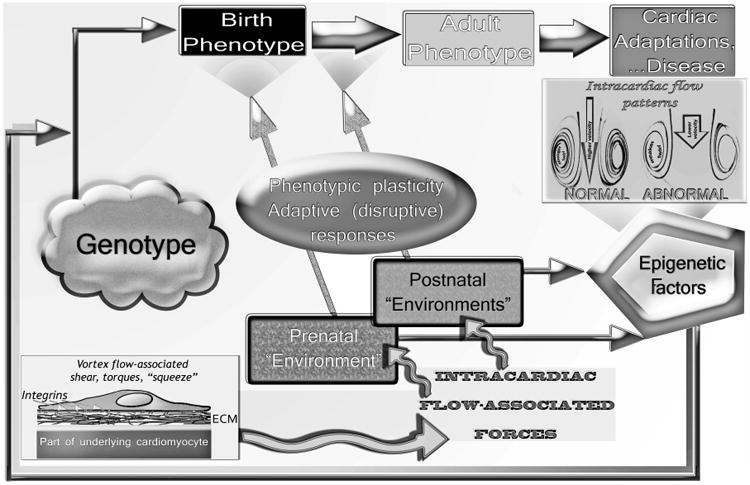
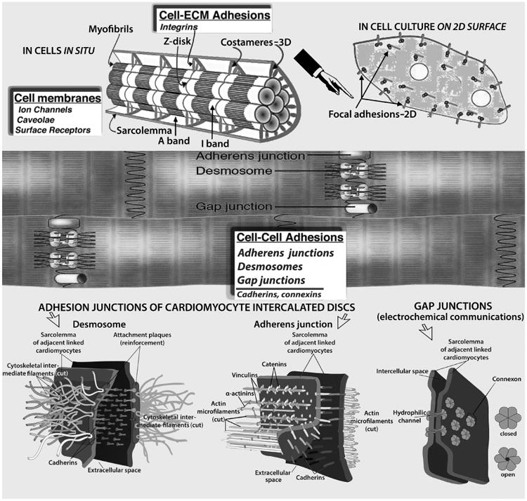
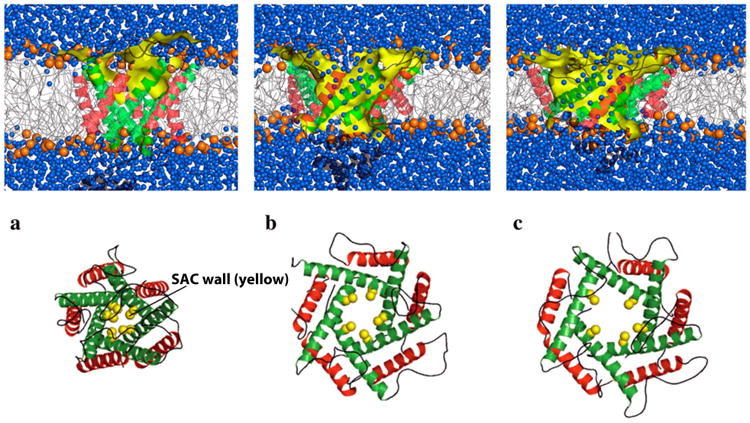
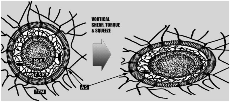
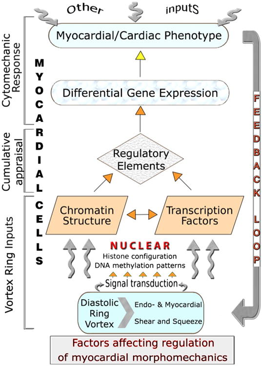
Epigenetic mechanisms are fundamental in cardiac adaptations, remodeling, reverse remodeling, and disease. A primary goal of translational cardiovascular research is recognizing whether disease-related changes in phenotype can be averted by eliminating or reducing the effects of environmental epigenetic risks. There may be significant medical benefits in using gene-by-environment interaction knowledge to prevent or reverse organ abnormalities and disease. This survey proposes that "environmental" forces associated with diastolic RV/LV rotatory flows exert important, albeit still unappreciated, epigenetic actions influencing functional and morphological cardiac adaptations. Mechanisms analogous to Murray's law of hydrodynamic shear-induced endothelial cell modulation of vascular geometry are likely to link diastolic vortex-associated shear, torque and "squeeze" forces to RV/LV adaptations. The time has come to explore a new paradigm in which such forces play a fundamental epigenetic role, and to work out how heart cells react to them. Findings from various imaging modalities, computational fluid dynamics, molecular cell biology and cytomechanics are considered. The following are examined, among others: structural dynamics of myocardial cells (endocardium, cardiomyocytes, and fibroblasts), cytoskeleton, nucleoskeleton, and extracellular matrix; mechanotransduction and signaling; and mechanical epigenetic influences on genetic expression. To help integrate and focus relevant pluridisciplinary research, rotatory RV/LV filling flow is placed within a working context that has a cytomechanics perspective. This new frontier in cardiac research should uncover versatile mechanistic insights linking filling vortex patterns and attendant forces to variable expressions of gene regulation in RV/LV myocardium. In due course, it should reveal intrinsic homeostatic arrangements that support ventricular myocardial function and adaptability.


Figures

Figure 1
Figure 1. Genotype × Environment = Phenotype
A circular regulatory pathway exists during cardiogenesis and in pre- and postnatal life between: the intracardiac blood flow patterns and associated shear, torque, and “squeeze” (see discussion in text) force transmission to the myocardial walls; epigenetically influenced gene expression; and changes in the morphology of the developing prenatal or in the phenotype of the continuously adapting postnatal and adult heart. Phenotypic plasticity can, in conjunction with normal and anomalous operating “environmental” conditions, lead not only to adaptive but also to disruptive (maladaptive) responses, morphomechanical cardiac abnormalities and disease. Slightly modified with permission of PMPH-USA from Pasipoularides A. Heart's Vortex: Intracardiac Blood Flow Phenomena. Shelton, CT: People's Medical Publishing House, 2010. 960 pp.
Figure 2
Figure 2
Myocardial cell junctions can be divided into two types: those that link cells to the ECM (costameres and focal adhesions, Top), and intercellular junctions (gap, adherens, and desmosomes, Middle & Bottom) that link cells together directly. Radially arranged integrins, and other specialized proteins, constitute physical links between the Z-disk and sarcolemma; they transmit contractile forces from sarcomeres across the sarcolemma laterally to the extracellular matrix and ultimately to neighboring cardiomyocytes. The desmosome, like the adherens junction, comprises calcium ion-dependent cell adhesion molecules that interact with similar molecules in the adjacent cell. Adherens junctions and focal adhesions not only tether cells together or to the ECM, but they also transduce signals into and out of the cell, influencing a variety of cellular epigenetic responses, notably to flow-associated forces and deformations. The basic building block of the gap junction is the connexin subunit. Six of these in each of the membranes of two adjacent cells come together to form a connexon that interacts with a comparable hexamer in the other cell resulting in formation of a channel, which allows cytoplasmic communication between the cells. Myocardial cell membranes encompass ion channels, surface receptors and caveolae—not shown. (See discussions in text). Costamere diagram adapted, modified, from Ervasti JM [31].
Figure 3
Figure 3
Schematic of numerical simulation of the gating process of a sarcolemmal mechanosensitive ion channel. Putting the membrane under tension, the stretch-activated channels (SAC) undergo significant conformational changes in accordance with an iris-like dilation mechanism, reaching a conducting state on a microsecond timescale. Diagrammed is a channel with (a) small, (b) intermediate, and (c) large opening under the action of rising membrane tension. Top: side view showing the lipid bilayer (blue denotes an aqueous environment, yellow represents the SAC wall). Bottom: End view showing the simulated membrane pore. Reproduced, slightly modified, with permission from Yefimov S, et al [77].
Figure 4
Figure 4
Schematic depiction of the global mechanical interconnectedness of the myocardial extracellular matrix (ECM), cytoskeleton (CSK), and nucleoskeleton (NSK), which has a pivotal role in the epigenetic dynamic actions of the right and left ventricular diastolic toroidal vortex. The inner and outer nuclear membranes and lamins anchored to the nuclear envelope are demarcated. The ECM, CSK, and NSK form a continuous and interconnected communication system, all parts of which work together synergistically. Adhesion structures (AS) are spatially discrete, transmembrane cadherin (cell–cell adhesions) and integrin (cell–ECM adhesions) complexes; they activate many of the same signaling pathways and provoke similar cellular activities, acting as interdependent functional nodes in a larger cytomechanics network. Through the intermediation of ECM, CSK and NSK structures, extra- and intra-cellular forces alter nuclear shape, promote nucleosome disruption and chromatin relaxation, and alter gene activities. (See discussion in text).
Figure 5
Figure 5
Summary of epigenetic dynamic actions of RV/LV diastolic toroidal vortex: Cytoskeletal “tensegrity” dynamics create equilibrium between strut compression and string tension, allowing internal structural balance and shape maintenance. Cells sense their physical 3D “environment,” including variable diastolic vortical shear, torque and “squeeze” forces, by transducing mechanical deformations and forces into differentiated transcription and translation signals, which can adjust cellular and extracellular tissue and organ phenotype; the latter, in turn, affects (feedback loop) diastolic filling vortex strength and vortical shear and “squeeze” forces. Mechanosensitive regulatory controls modulate myocyte shape and intracellular architecture, and processes as diverse as proliferation, hypertrophy, and apoptosis, involved in cardiac homeostasis and adaptations.

Similar articles

Cited by

References

    1. Pasipoularides A. Heart's Vortex: Intracardiac Blood Flow Phenomena. Shelton, CT: People's Medical Publishing House; 2010. p. 960.
    1. Pasipoularides A. Diastolic filling vortex forces and cardiac adaptations: probing the epigenetic nexus. Hellenic J Cardiol. 2012;53:458–69. - PMC - PubMed
    1. Pasipoularides A. Mechanotransduction mechanisms for intraventricular diastolic vortex forces and myocardial deformations: Part 1. J Cardiovasc Transl Res. 2015;8:76–87. doi: 10.1007/s12265-015-9611-y. - DOI - PMC - PubMed
    1. Hines LM, Stampfer MJ, Ma J, et al. Genetic variation in alcohol dehydrogenase and the beneficial effect of moderate alcohol consumption on myocardial infarction. N Engl J Med. 2001;344:549–555. - PubMed
    1. Talmud PJ, Stephens JW, Hawe E, et al. The significant increase in cardiovascular disease risk in APOEepsilon4 carriers is evident only in men who smoke: potential relationship between reduced antioxidant status and ApoE4. Ann Hum Genet. 2005;69:613–622. - PubMed

Publication types

Substances

LinkOut - more resources