. 2015 Jun;37(3):9792.
doi: 10.1007/s11357-015-9792-y. Epub 2015 May 14.

Development of monosodium acetate-induced osteoarthritis and inflammatory pain in ageing mice

Affiliations

Development of monosodium acetate-induced osteoarthritis and inflammatory pain in ageing mice

Andrea C Ogbonna et al. Age (Dordr). 2015 Jun.

Abstract

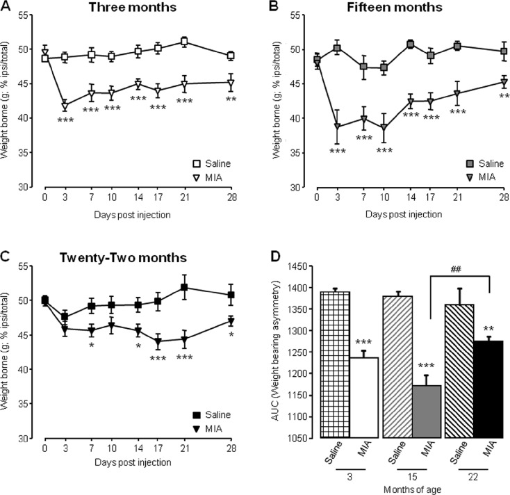
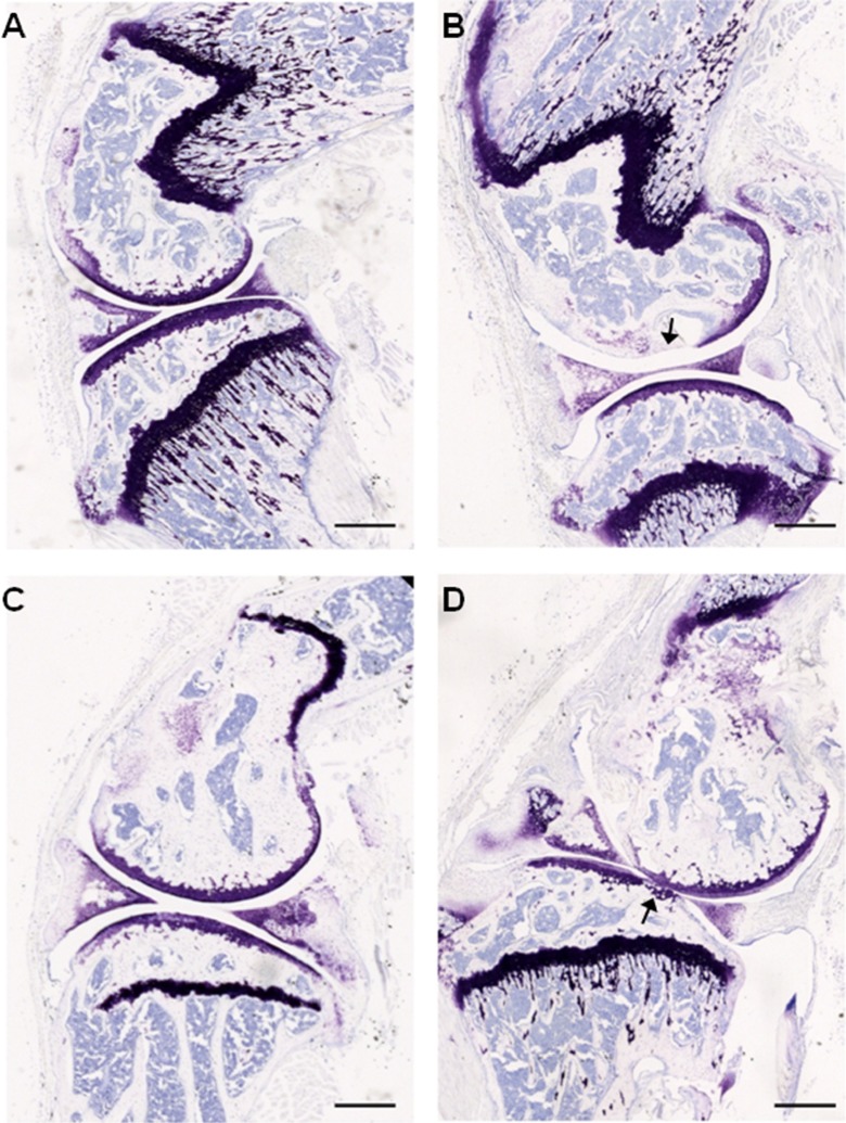
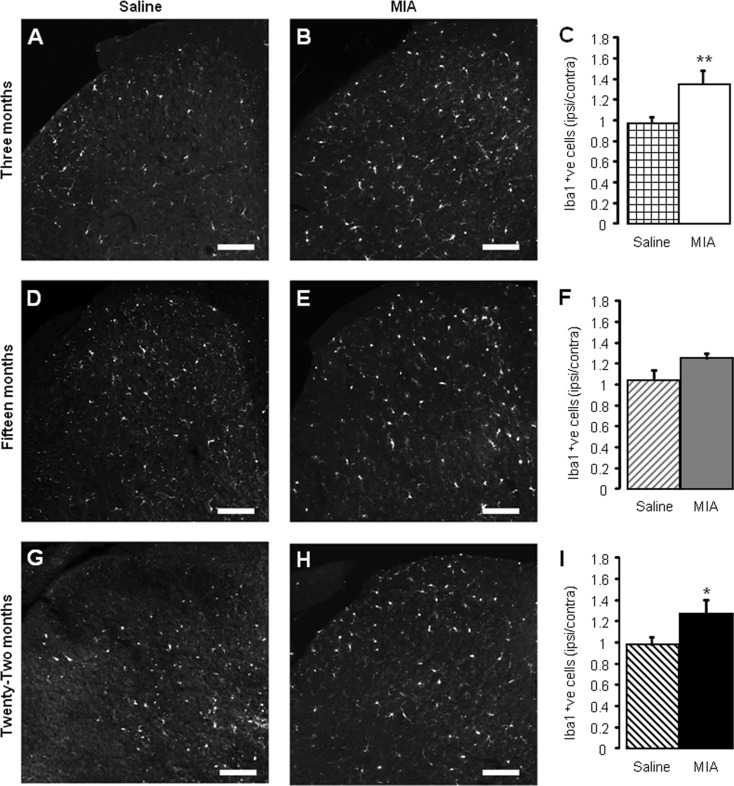
Most conditions associated with ageing result from an age-related loss in the function of cells and tissues that maintain body homeostasis. In osteoarthritis (OA) patients, an inadequate response to stress or joint injury can lead to tissue destruction which can result in chronic pain. Here, we evaluated the development of monoiodoacetate (MIA)-induced OA in 3-, 15- and 22-month-old mice and assessed the pain-like behaviours and the spinal microglial changes associated with MIA administration. We observed that in aged mice, nocifensive behaviour was significantly attenuated in comparison to young adults despite similar knee joint pathology. Specifically referred mechanical allodynia associated with the MIA initial inflammatory phase (0-10 days) was significantly attenuated in 22-month-old mice. In contrast, the late phase of MIA-induced mechanical allodynia was comparable between age groups. Significant increase of microglia cell numbers was detected in 3, but not 15- and 22-month-old spinal cords. Furthermore, in the zymosan model of acute inflammation, mechanical allodynia was attenuated, and microglial response was less robust in 22 compared to 3-month-old mice. This study suggests that nocifensive responses to damaging stimuli are altered with advancing age and microglial response to peripheral damage is less robust.


Figures

Fig. 1
Fig. 1
Ageing affects MIA-associated weight bearing asymmetry. ac Weight borne on ipsilateral hind limb was monitored at regular intervals in 3-month (a), 15-month (b) and 22-month (c)-old mice before and until 28 days following intra-articular injection. ***p < 0.001, **p < 0.01, *p < 0.05 compared to saline control group, two-way RM ANOVA post hoc Student Newman-Keuls. d Area under the curve (AUC) analysis of weight borne ***p < 0.001, **p < 0.01 compared to age-matched saline control; ##p < 0.01 between 15- and 22-month-old mice; two-way ANOVA, post hoc Student Newman-Keuls. Data shown as mean ± SEM, n = 10–16 mice per group
Fig. 2
Fig. 2
Ageing affects MIA-associated referred mechanical hypersensitivity. ac Mechanical withdrawal responses of ipsilateral hind paws were measured before and at regular intervals until 28-day post-intra-articular injection in 3-month (a), 15-month (b) and 22-month (c)-old mice. White bars represent the duration of the early and late phases of mechanical hypersensitivity. ***p < 0.001, **p < 0.01 compared to saline control group, two-way RM ANOVA, post hoc Student Newman-Keuls (de); AUC was calculated for all groups from 0 to 10 days (early phase; d) and from 14 to 28 days (late phase; e). ***p < 0.001 compared to age-matched saline control group, ## p < 0.01; #p < 0.05 between groups as indicated, two-way ANOVA, post hoc Student Newman-Keuls. Data are shown as mean ± SEM, n = 10–16 mice per group
Fig. 3
Fig. 3
Intra-articular MIA administration results in knee joint pathology in young and aged mice. Toluidine blue staining of articular cartilage from saline (a, c) and MIA (b, d) injected ipsilateral knee joints. Smooth and intact articular surface of the femur and tibia from 3-month (a) and 22-month (c)-old mice injected with saline in contrast to thinning and loss of articular integrity (indicated by arrows) in 3-month (b) and 22-month (d)-old mice injected with 1-mg MIA. Scale bar = 500 μm
Fig. 4
Fig. 4
MIA-associated microglial response is attenuated in aged mice. Iba-1 immunoreactivity in L5 ipsilateral dorsal horn in saline (a, d, g) and MIA-treated (b, e, h) mice 28 days after intra-articular administration in 3- (a, b), 15- (d, e) and 22- (g, h) month-old mice. Scale bars = 100 μm. Quantification of Iba-1 immunoreactivity in saline and MIA-treated dorsal horn 28 days after administration in 3- (c), 15- (f) and 22- (i) month-old mice. Data shown as mean ± SEM, **p < 0.01, *p < 0.05 compared to age-matched saline group, two-way ANOVA, post hoc Student Newman-Keuls, n = 4–5 spinal cords per group
Fig. 5
Fig. 5
Zymosan-induced nocifensive behaviour and microglial response are attenuated in aged mice (ab); mechanical withdrawal responses (a) and thermal withdrawal latencies (b) of ipsilateral hind paws were measured before (pre) and 24-h post-intra-plantar injection of zymosan (Zym; 20 μL; 0.2 mg/mL). Data shown as mean ± SEM, ***p < 0.001, **p < 0.01, compared to baseline (pre) threshold, two-way ANOVA, post hoc Student Newman-Keuls, n = 5 mice per group. c Quantification of Iba-1 immunoreactivity (per 104 μm2) in ipsilateral and contralateral lumbar (L4 and L5) dorsal horn 24 h after intraplantar zymosan administration (Zym; 20 μL; 0.2 mg/mL) compared to naive dorsal horn from age-matched controls. d Quantification of ipsilateral Iba-1 immunoreactivity is normalised to contralateral levels. Data shown as mean ± SEM, **p < 0.01, *p < 0.05, compared to age-matched naive, #p < 0.05 compared to indicated group, two-way ANOVA post hoc Student Newman-Keuls, n = 5 spinal cords per group

Similar articles

Cited by

References

    1. Bove SE, Calcaterra SL, Brooker RM, Huber CM, Guzman RE, Juneau PL, Schrier DJ, Kilgore KS. Weight bearing as a measure of disease progression and efficacy of anti-inflammatory compounds in a model of monosodium iodoacetate-induced osteoarthritis. Osteoarthr Cartil. 2003;11:821–830. doi: 10.1016/S1063-4584(03)00163-8. - DOI - PubMed
    1. Ceballos D, Cuadras J, Verdu E, Navarro X. Morphometric and ultrastructural changes with ageing in mouse peripheral nerve. J Anat. 1999;195:563–576. doi: 10.1046/j.1469-7580.1999.19540563.x. - DOI - PMC - PubMed
    1. Chaplan SR, Bach FW, Pogrel JW, Chung JM, Yaksh TL. Quantitative assessment of tactile allodynia in the rat paw. J Neurosci Methods. 1994;53:55–63. doi: 10.1016/0165-0270(94)90144-9. - DOI - PubMed
    1. Clark AK, Gentry C, Bradbury EJ, McMahon SB, Malcangio M. Role of spinal microglia in rat models of peripheral nerve injury and inflammation. Eur J Pain. 2007;11:223–230. doi: 10.1016/j.ejpain.2006.02.003. - DOI - PubMed
    1. Clark AK, Grist J, Al-Kashi A, Perretti M, Malcangio M. Spinal cathepsin S and fractalkine contribute to chronic pain in the collagen-induced arthritis model. Arthritis Rheum. 2012;64:2038–2047. doi: 10.1002/art.34351. - DOI - PubMed

Substances

LinkOut - more resources