. 2015 May 14:14:103.
doi: 10.1186/s12943-015-0367-4.

Quercetin inhibits HGF/c-Met signaling and HGF-stimulated melanoma cell migration and invasion

Affiliations

Quercetin inhibits HGF/c-Met signaling and HGF-stimulated melanoma cell migration and invasion

Hui-Hui Cao et al. Mol Cancer. .

Abstract

Background: Melanoma is notorious for its propensity to metastasize, which makes treatment extremely difficult. Receptor tyrosine kinase c-Met is activated in human melanoma and is involved in melanoma progression and metastasis. Hepatocyte growth factor (HGF)-mediated activation of c-Met signaling has been suggested as a therapeutic target for melanoma metastasis. Quercetin is a dietary flavonoid that exerts anti-metastatic effect in various types of cancer including melanoma. In a previous report, we demonstrated that quercetin inhibited melanoma cell migration and invasion in vitro, and prevented melanoma cell lung metastasis in vivo. In this study, we sought to determine the involvement of HGF/c-Met signaling in the anti-metastatic action of quercetin in melanoma.

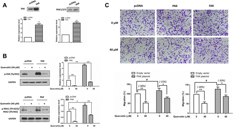
Methods: Transwell chamber assay was conducted to determine the cell migratory and invasive abilities. Western blotting was performed to determine the expression levels and activities of c-Met and its downstream molecules. And immunoblotting was performed in BS(3) cross-linked cells to examine the homo-dimerization of c-Met. Quantitative real-time PCR analysis was carried out to evaluate the mRNA expression level of HGF. Transient transfection was used to overexpress PAK or FAK in cell models. Student's t-test was used in analyzing differences between two groups.

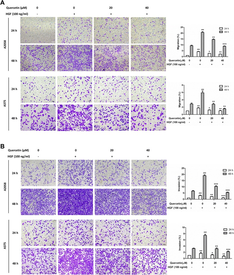
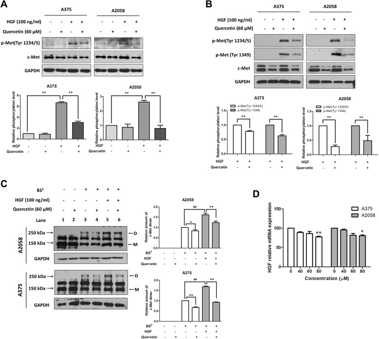
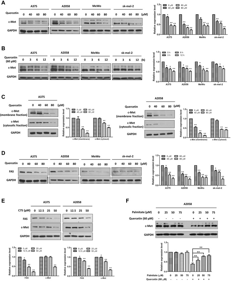
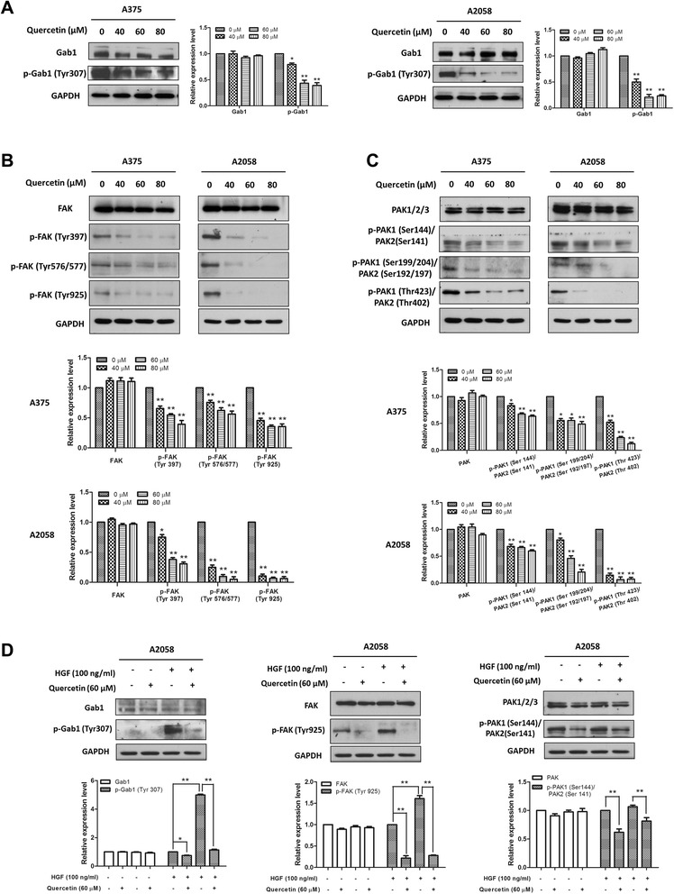
Results: Quercetin dose-dependently suppressed HGF-stimulated melanoma cell migration and invasion. Further study indicated that quercetin inhibited c-Met phosphorylation, reduced c-Met homo-dimerization and decreased c-Met protein expression. The effect of quercetin on c-Met expression was associated with a reduced expression of fatty acid synthase. In addition, quercetin suppressed the phosphorylation of c-Met downstream molecules including Gab1 (GRB2-associated-binding protein 1), FAK (Focal Adhesion Kinase) and PAK (p21-activated kinases). More importantly, overexpression of FAK or PAK significantly reduced the inhibitory effect of quercetin on the migration of the melanoma cells.

Conclusions: Our findings suggest that suppression of the HGF/c-Met signaling pathway contributes to the anti-metastatic action of quercetin in melanoma.


Figures

Figure 1
Figure 1
Quercetin suppressed HGF-stimulated melanoma cell migration and invasion. A2058 and A375 cells were seeded onto the upper chamber consisting of 8 μm pore-size filters coated without (A) and with (B) Matrigel basement membrane matrix, then treated with indicated concentration of quercetin for 24 h or 48 h without (−) or with (+) 100 ng/ml HGF in the lower chamber. The relative quantitative determinations of migrated and invasive cells were calculated with 5 fields counted per experiment. Data were shown as mean ± S.D. from three independent experiments, ### P < 0.001 compared with unstimulated and untreated cells, *P < 0.05, **P < 0.01 and ***P < 0.001 compared with stimulated and untreated cells.
Figure 2
Figure 2
Quercetin inhibited the activation of c-Met in melanoma cells. (A) Cells were treated with the indicated concentrations of quercetin for 6 h and then stimulated with or without HGF (100 ng/ml) for 10 min. Levels of c-Met and phospho-Met in total lysates were evaluated by immunoblotting. The representative results (upper) and the relative phosphorylation levels (bottom) were shown. The relative levels of phospho-Met were calculated after normalizing the levels of phospho-Met to total c-Met. (B) Cells were grown in serum-free medium overnight before incubation with the indicated concentrations of quercetin for 6 h, after stimulation with or without 100 ng/ml HGF for 10 min, cells were lysed for immunoblotting. The representative results (upper) and the relative phosphorylation levels (bottom) were shown. The relative levels of phospho-Met were calculated after normalizing the levels of phospho-Met to the total c-Met. (C) Quercetin reduced c-Met homo-dimerization. Cells were starved overnight and then treated with indicated concentrations of quercetin for 6 h. After that, these cells were stimulated with or without HGF (100 ng/ml) for 10 min, and then incubated with BS3. Cell lysates were prepared for immunoblotting by using an anti-c-Met antibody. The arrows indicated the dimer (D) and the monomer (M) c-Met. Molecular weights of the marker are shown on the left. The representative results (left) and the relative expression levels of dimer c-Met (right) were shown. The relative levels of dimer c-Met were analyzed after normalizing the levels of dimer c-Met to monomer c-Met. (D) Effect of quercetin on HGF mRNA expression. Cells were treated with indicated concentrations of quercetin for 24 h and then the real-time PCR analysis was performed to detect the mRNA expression levels of HGF. Data were mean ± S.D. from three independent experiments. *P < 0.05, **P < 0.01 and ## P < 0.01.
Figure 3
Figure 3
Quercetin reduced c-Met levels through the inhibition of FAS. A375, A2058, MeWo and sk-mel-2 cells were treated with (A) various concentrations of quercetin for 24 h or (B) a fixed concentration (60 μM) for various durations, and then the whole-cell lysates were prepared and the Western blot analysis was performed to determine the expression level of c-Met. (C) A375 and A2058 cells were treated with indicated concentrations of quercetin for 24 h, and then the membrane and cytosolic lysates were prepared. The expression levels of c-Met in the two fractions were examined by immunoblotting. (D) A375, A2058, MeWo and sk-mel-2 cells were treated with indicated concentrations of quercetin for 24 h, and then the expression of FAS were determined by immunoblotting. (E) A375 and A2058 cells were treated with indicated concentrations of C75 for 24 h and then the immunoblotting assay was conducted to determine the expression levels of FAS and c-Met. (F) A2058 cells were exposed to either the vehicle or 60 μM quercetin for 24 h in the absence or presence of palmitate, and then the whole-cell lysates were prepared for immunoblotting by using a c-Met antibody. Independent experiments were performed at least three times, and the results from a representative experiment are shown. The relative expression levels were analyzed by Image J software and shown as mean ± S.D., *P < 0.05, **P < 0.01.
Figure 4
Figure 4
Quercetin suppressed the activation of c-Met downstream molecules. (A, B and C) A375 and A2058 cells were treated with indicated concentrations of quercetin for 24 h. (D) A2058 cells were starved overnight and then exposed to the vehicle or quercetin (60 μM) for 6 h. After that, these cells were stimulated with or without HGF (100 ng/ml) for 10 min. For each experiment, the whole-cell lysates were prepared and probed by Western blot using specific antibodies. Independent experiments were performed at least three times, and the results from a representative experiment are shown. The relative expression levels were analyzed by Image J software and shown as mean ± S.D., *P < 0.05, **P < 0.01.
Figure 5
Figure 5
Overexpression of PAK or FAK partially reversed the migration-inhibition effect of quercetin. A2058 cells were transiently transfected with a FAK-expressing construct, a PAK-expressing construct, or an empty vector for 24 h. (A) Expression levels of FAK (or PAK) on cells that transfected with FAK (or PAK)-expressing construct. (B) After transfection, cells were treated with quercetin for 24 h, and the effects of quercetin on FAK (or PAK) overexpressed cells were determined by immunoblotting. (C) Cell migratory abilities were measured by migration chamber assay after treated with quercetin for 48 h. The relative quantitative determinations of migrated cells were calculated with 5 fields counted per experiment. The data shown here are the mean ± S.D. from three independent experiments. # P < 0.05, *P < 0.05 and ** < 0.01.

Similar articles

Cited by

References

    1. Siegel R, DeSantis C, Virgo K, Stein K, Mariotto A, Smith T, et al. Cancer treatment and survivorship statistics, 2012. CA Cancer J Clin. 2012;62:220–241. doi: 10.3322/caac.21149. - DOI - PubMed
    1. Cummins DL, Cummins JM, Pantle H, Silverman MA, Leonard AL, Chanmugam A. Cutaneous malignant melanoma. Mayo Clin Proc. 2006;81:500–507. doi: 10.4065/81.4.500. - DOI - PubMed
    1. Serrone L, Zeuli M, Sega FM, Cognetti F. Dacarbazine-based chemotherapy for metastatic melanoma: thirty-year experience overview. J Exp Clin Cancer Res. 2000;19:21–34. - PubMed
    1. Alwan LM, Grossmann K, Sageser D, Van Atta J, Agarwal N, Gilreath JA. Comparison of acute toxicity and mortality after two different dosing regimens of high-dose interleukin-2 for patients with metastatic melanoma. Target Oncol. 2014;9:63–71. doi: 10.1007/s11523-013-0276-7. - DOI - PubMed
    1. Atkins MB, Lotze MT, Dutcher JP, Fisher RI, Weiss G, Margolin K, et al. High-dose recombinant interleukin 2 therapy for patients with metastatic melanoma: analysis of 270 patients treated between 1985 and 1993. J Clin Oncol. 1999;17:2105–2116. - PubMed

Publication types

MeSH terms