. 2015 Jul;34(3):195-205.
doi: 10.14366/usg.15006. Epub 2015 Mar 10.

Postoperative ultrasonography of the musculoskeletal system

Affiliations

Postoperative ultrasonography of the musculoskeletal system

Kyung Ah Chun et al. Ultrasonography. 2015 Jul.

Abstract

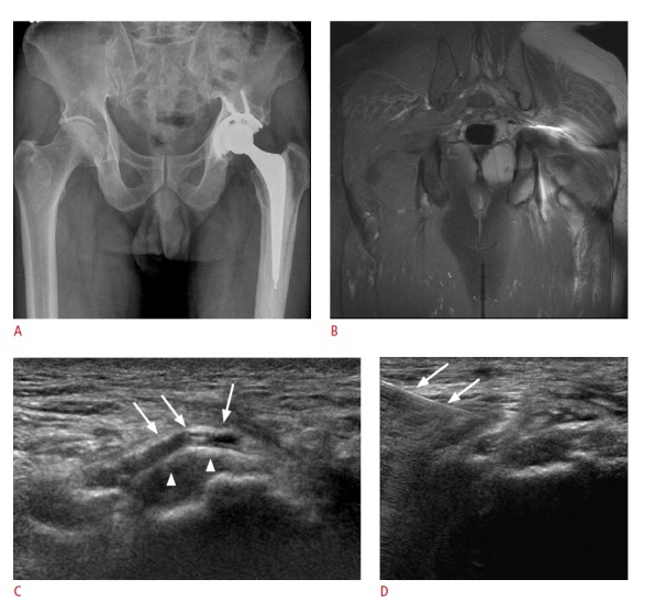
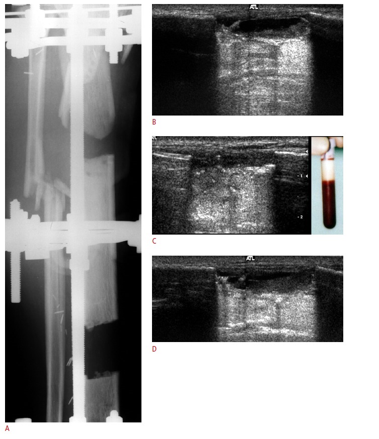
Ultrasonography of the postoperative musculoskeletal system plays an important role in the accurate diagnosis of abnormal lesions in the bone and soft tissues. Ultrasonography is a fast and reliable method with no harmful irradiation for the evaluation of postoperative musculoskeletal complications. In particular, it is not affected by the excessive metal artifacts that appear on computed tomography or magnetic resonance imaging. Another benefit of ultrasonography is its capability to dynamically assess the pathologic movement in joints, muscles, or tendons. This article discusses the frequent applications of musculoskeletal ultrasonography in various postoperative situations including those involving the soft tissues around the metal hardware, arthroplasty, postoperative tendons, recurrent soft tissue tumors, bone unions, and amputation surgery.

Keywords: Musculoskeletal system; Postoperative care; Soft tissue infections; Soft tissue injuries; Ultrasonography.


Conflict of interest statement

No potential conflict of interest relevant to this article was reported.

Figures

Fig. 1.
Fig. 1.. A 32-year-old woman with a postoperative infection after intramedullary nailing.
A. Oblique radiograph shows suspicious cortical lucency along the medial aspect of the distal tibia. B. Longitudinal sonogram at the distal tibia shows subperiosteal anechoic abscesses (arrows) with a hyperechoic nail and screw. C. Postcontrast computed tomography shows irregularly shaped, non-enhancing hypodense soft tissue lesions (arrows) just medial to the tibia with subcutaneous edema. The abscess was surgically confirmed.
Fig. 2.
Fig. 2.. A 66-year-old woman with a previous comminuted fracture of the elbow treated with surgical pins.
A, B. Postoperative sonogram (A) shows a large elongated hypoechoic lesion (arrowheads) adjacent to the distal humerus with a hyperechoic pin (arrow) and a reverberation artifact, suggesting a hematoma due to the orthopedic K-wire (arrow) noted on the radiograph (B). The blood was surgically drained.
Fig. 3.
Fig. 3.. A 74-year-old woman with an infection 10 months after hemiarthroplasty.
A. The anterior-posterior radiograph shows the hemiarthroplasty of the left hip with soft tissue swelling. B. Sonogram of the proximal femur shows a subperiosteal hypoechoic lesion and an anechoic abscess containing debris (arrows).
Fig. 4.
Fig. 4.. A 72-year-old man with pain in the distal thigh after internal fixation.
A. The anterior-posterior radiograph shows internal fixation along the left femur and soft tissue swelling of the distal thigh. B. Sonogram of the distal thigh shows hyperechoic screws (arrows) of the distal femur and an adjacent irregular anechoic lesion with a thick wall, representing the abscess. C. The color Doppler sonogram shows a perilesionally increased vascular flow. Staphylococcus aureus grew on the culture plate.
Fig. 5.
Fig. 5.. A 69-year-old man with left buttock pain after total hip arthroplasty.
A. The anterior-posterior radiograph shows total hip arthoplasty with no definite abnormality. B. Magnetic resonance image shows poor delineation of structures due to the artifact. C. Sonogram shows anechoic fluid collection (arrows) just posterior to ischial tuberosity (arrowheads), suggesting ischio-gluteal bursitis. D. Ultrasound-guided steroid injection (arrows) into the bursa was performed.
Fig. 6.
Fig. 6.. A 54-year-old man with a supraspinatus tendon 6 months after repair.
Sonogram shows heterogeneous echo texture in the repaired tendon. Further, surface irregularity and small curvilinear echogenicity (arrows) along the anchor of the greater tuberosity with the suture material (arrowheads) within the tendon were noted.
Fig. 7.
Fig. 7.. A 51-year-old woman with a supraspinatus tendon 6 months after repair.
Sonogram shows a normal fibrillar pattern of the repaired tendon (arrowheads) and an anchor with a reverberation artifact (arrows).
Fig. 8.
Fig. 8.. A 51-year-old man with a supraspinatus tendon 6 months after repair.
Sonogram shows a heterogeneous echogenicity without a fibrillar pattern along the repaired tendon and a small focal low echoic area (arrowhead) representing the granulation tissue. Peribursal thickening is also seen (arrows). The patient exhibited no shoulder symptoms after tendon repair.
Fig. 9.
Fig. 9.. A 73-year-old man with a recurrent tear of the repaired tendon.
Sonogram shows a large irregular defect with anechoic fluid collection (arrowheads) in the repaired tendon, and broken suture (arrow).
Fig. 10.
Fig. 10.. A 63-year-old man with a massive retear of the repaired tendon.
A. Sonogram shows the absence of the repaired tendon with extensive anechoic fluid collection (arrows). B. The corresponding T2-weighted magnetic resonance image shows the absence of the repaired tendon with hyperintense fluid collection (arrowheads).
Fig. 11.
Fig. 11.. A 37-year-old man with a retear of the Achilles tendon 4 months after repair.
A. Sonogram shows an irregular anechoic lesion (arrowheads) in the markedly thickened repaired tendon and the displaced suture material (arrow). B. The color Doppler sonogram shows an increased vascular flow along the repaired tendon with marked thickening.
Fig. 12.
Fig. 12.. A 45-year-old man with a postoperative mass palpated in the area of surgery after resection.
Sonogram shows anechoic fluid collection along the muscle fascia, suggesting a seroma.
Fig. 13.
Fig. 13.. A 52-year-old man with bone lengthening following the Ilizarov procedure.
A. The anterior-posterior radiograph shows multiple bone defects with the Ilizarov external fixator. B. Sonogram shows an anechoic fluid lesion in the osteotomy defect. C. The blood was aspirated under ultrasound-guidance, indicating a hematoma. D. Ultrasonography conducted 2 weeks after aspiration showed considerable regression of the hematoma from 1.8 cm to 0.8 cm.
Fig. 14.
Fig. 14.. A 47-year-old man with tingling pain in the amputation stump.
A. The anterior-posterior radiograph shows the status of the amputation below the knee. B. Sonogram shows a hypoechoic mass (arrow) in continuity with the peroneal nerve (arrowheads). A neuroma was confirmed by surgical resection.

References

    1. Jacobson JA. Musculoskeletal sonography and MR imaging: a role for both imaging methods. Radiol Clin North Am. 1999;37:713–735. - PubMed
    1. Jacobson JA, Lax MJ. Musculoskeletal sonography of the postoperative orthopedic patient. Semin Musculoskelet Radiol. 2002;6:67–77. - PubMed
    1. Guillin R, Botchu R, Bianchi S. Sonography of orthopedic hardware impingement of the extremities. J Ultrasound Med. 2012;31:1457–1463. - PubMed
    1. Sofka CM, Adler RS. Original report. Sonographic evaluation of shoulder arthroplasty. AJR Am J Roentgenol. 2003;180:1117–1120. - PubMed
    1. van Holsbeeck MT, Eyler WR, Sherman LS, Lombardi TJ, Mezger E, Verner JJ, et al. Detection of infection in loosened hip prostheses: efficacy of sonography. AJR Am J Roentgenol. 1994;163:381–384. - PubMed

LinkOut - more resources