. 2015 Jul 17;290(29):17796-17805.
doi: 10.1074/jbc.M114.617613. Epub 2015 May 13.

Computation and Functional Studies Provide a Model for the Structure of the Zinc Transporter hZIP4

Affiliations

Computation and Functional Studies Provide a Model for the Structure of the Zinc Transporter hZIP4

Sagar Antala et al. J Biol Chem. .

Abstract

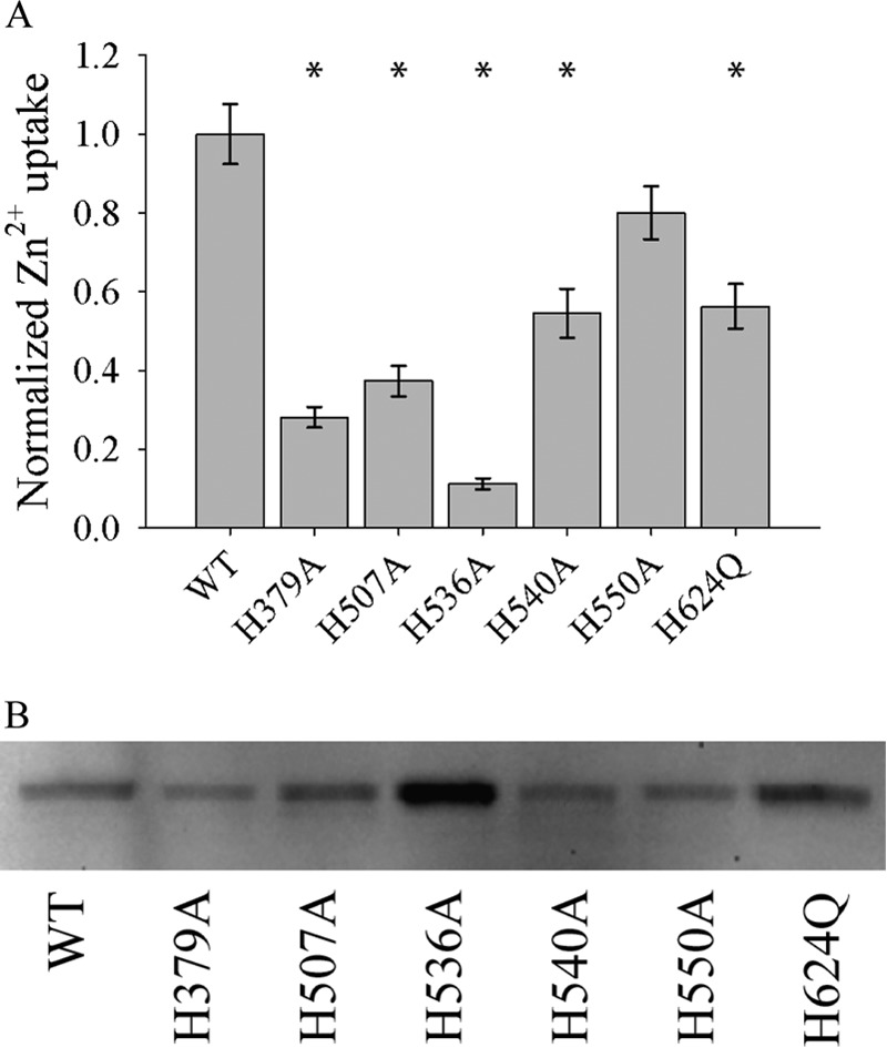
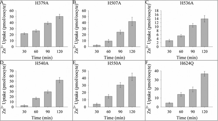
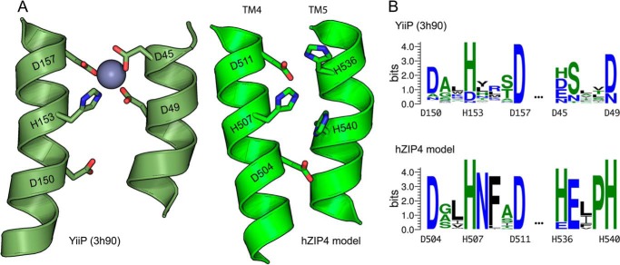
Members of the Zrt and Irt protein (ZIP) family are a central participant in transition metal homeostasis as they function to increase the cytosolic concentration of zinc and/or iron. However, the lack of a crystal structure hinders elucidation of the molecular mechanism of ZIP proteins. Here, we employed GREMLIN, a co-evolution-based contact prediction approach in conjunction with the Rosetta structure prediction program to construct a structural model of the human (h) ZIP4 transporter. The predicted contact data are best fit by modeling hZIP4 as a dimer. Mutagenesis of residues that comprise a central putative hZIP4 transmembrane transition metal coordination site in the structural model alter the kinetics and specificity of hZIP4. Comparison of the hZIP4 dimer model to all known membrane protein structures identifies the 12-transmembrane monomeric Piriformospora indica phosphate transporter (PiPT), a member of the major facilitator superfamily (MFS), as a likely structural homolog.

Keywords: computer modeling; membrane biophysics; membrane protein; metal homeostasis; metal ion-protein interaction; protein evolution; transport metal; transporter; zinc.


Figures

FIGURE 1.
FIGURE 1.
A, sigmoidal distance restraint function. d is the distance between given distance evaluated. B, strong repulsive distance restraints were added between extracellular regions and intracellular regions, and strong attractive restraints were added within intracellular regions and extracellular regions, effectively constructing a membrane-like sampling space.
FIGURE 2.
FIGURE 2.
A, contact map, left to right, top to bottom (N to C terminus) showing the top 384 co-evolving residues between transmembrane helices represented as a contact map. The darker and larger the blue dots, the higher strength in covariance. B, table of interhelical residue pairs, within the top 64 predictions. The matching labels are shown in the bottom left portion of the contact map (A).
FIGURE 3.
FIGURE 3.
Distribution of GREMLIN restraint scores for Rosetta ab initio models. Models above the red line (with z-score ≥2) were selected for the clustering analysis. The selected model is the refined model from cluster A (Fig. 4A). The ab initio models closer to the selected model tend to have a higher GREMLIN restraint score.
FIGURE 4.
FIGURE 4.
Contact maps showing contacts made within each cluster (top) and cartoons of the top three models colored blue to red (N to C terminus) for each cluster (bottom). The intensity of the blue (in the contact map) is indicative of the percentage of models within the cluster that make those contacts. Intensity of blue ranges from light blue (10%) to blue (100%). x- and y-axis numbering (1–8) in contact maps represent transmembrane helices 1–8. Red boxes indicate helices predicted to be in contact but are not in the given cluster. A, cluster A makes contacts between all the top co-evolving helices, but there is a disagreement between models in the regions boxed in green. Further analysis reveals that variation within cluster A is explained within the context of dimer helix swapping (Fig. 6). B and C, both clusters B and C have missing helical contacts in regions with strong co-evolution signal. Additionally, they both expose histidines into the membrane, which is highly improbable for membrane proteins.
FIGURE 5.
FIGURE 5.
Models of hZIP4 using Rosetta structure prediction guided by co-evolution-based contacts. A, ribbon diagram of the monomer structure, colored blue to red (N to C terminus), shows the predicted location of the transmembrane histidines in white. For clarity, the loop between residues 428 and 486 is omitted. B, consistency of co-evolution predicted contacts with hZIP4 structural model. Black dots, predicted contacts; gray, contacts in the Rosetta model monomer; red, contacts across dimer interface. The top coevolving residues are made within the context of the dimer. x- and y-axis numbering (1–8) in the contact map represent transmembrane helices 1–8. C, dimer view of the Rosetta structure model of hZIP4. The top 25 contacts are shown as yellow dashes. The highlighted regions (red and dark blue) indicate the conserved LIV-1 sequence in the fourth transmembrane helix.
FIGURE 6.
FIGURE 6.
Variations within cluster A are consistent within context of a dimer. When we examine variation within cluster A, we find that the models are capable of making the remainder of the top co-evolving residues in the context of a dimer. For example, if we reconnect the loop regions between helices 2 and 3, swapping helix 3 between the homo-dimer (A ≥ B), this preserves all predicted helical contacts. Another example would be to reconnect the loop between helices 7 and 8, swapping helix 8 (A ≥ C), or a combination of both (A ≥ D). These swaps are represented in the variation of the top cluster (Fig. 4A).
FIGURE 7.
FIGURE 7.
Kinetics of H540A hZIP4 zinc transport. The transport parameters, Km and Vmax, of the H540A hZIP4 mutant were elucidated at low (A) and high (B) concentrations of zinc by measuring the amount of 65Zn2+ uptake into oocytes injected with hZIP4-Strep (with H540A mutant) mRNA over 1 h in assay buffer, which contained varying amounts of 65ZnCl2. The data were fit to the Michaelis-Menten equation described under “Materials and Methods.” Data originated from 4–7 oocytes; values are means ± S.E.
FIGURE 8.
FIGURE 8.
Relative velocity of WT hZIP4 and mutant transporters. A, oocytes were injected with 25 ng of WT hZIP4 or mutant hZIP4 mRNA. After 3 days of incubation at 16 °C, oocytes were subjected to radioisotope uptake experiment. Oocytes were incubated with 3 μm 65Zn2+, and zinc uptake was measured after 60 min. Transport data were normalized to surface expressed protein (B). * indicates a statistically significant difference in zinc uptake between mutant hZIP4 and WT after uptake was normalized to the level of protein surface expression (p < 0.05). Values are means ± S.E. B, Western blot of surface-expressed WT and mutant hZIP4 proteins isolated as described under “Materials and Methods.” The relative expression of hZIP4 was quantified using Quantity One software (Bio-Rad Laboratories).
FIGURE 9.
FIGURE 9.
Time course of zinc uptake for mutant constructs. Oocytes, injected with 25 ng of hZIP4 mutant constructs, were incubated with 21.75 μm 65ZnCl2. The assay was quenched every 30 min up to 120 min, and data originated from 5–8 oocytes; values are means ± S.E. Fitting the data to the equation to a standard linear equation (y = mx + b) resulted in the following R2 values: H379A (0.9685), H507A (0.9683), H536A (0.9824), H540A (0.9816), H550A (0.9949), and H624Q (0.9493).
FIGURE 10.
FIGURE 10.
Competition of 65Zn2+ uptake with a series of divalent cations. To determine which divalent cations inhibited hZIP4-mediated 65Zn2+ uptake, oocytes expressing hZIP4 were preincubated in 600 μm cold ZnCl2, BaCl2, CdCl2, CoCl2, CuCl2, FeCl2, MgCl2, MnCl2, or NiCl2 in the uptake assay buffer. The uptake assay was initiated by adding 3.0 μm 65ZnCl2. The assay was quenched after 1 h as described under “Materials and Methods.” Data were normalized to the amount of 65Zn2+ uptake in the absence of competing cation. Data originated from 14–17 oocytes; values are means ± S.E. Data marked with * represent statistically significant difference from WT (p < 0.05).
FIGURE 11.
FIGURE 11.
Structural homology of hZIP4 dimer to other membrane proteins. The red filled circles are MFS proteins. EmrE and YiiP are the efflux multidrug transporter and Znt zinc exporter, respectively.
FIGURE 12.
FIGURE 12.
A, similarity of the Rosetta model of hZIP4 with the structure of the MFS transporter (PDB: 4j05). Loop regions and helices 1 and 6 of hZIP4 are not shown for clarity. TM-score = 0.66 (3.53 root mean square deviation over aligned region). B, alignment of transmembrane helices in sequential space. The matching colors of hZIP4 and MFS indicate structural alignment. The gray regions did not align and were not shown in A.
FIGURE 13.
FIGURE 13.
Western blot analysis of tryptophan-scanning mutagenesis studies along TM7. Residues replaced with tryptophan are indicated above blot. Arrows represent (from top to bottom) dimer, glycosylated monomer, and nonglycosylated monomer.
FIGURE 14.
FIGURE 14.
A, expanded view of putative YiiP and hZIP4 transition metal coordination sites. B, WebLogo showing the conservation at each position.

References

    1. Eide D. J. (2004) The SLC39 family of metal ion transporters. Pflugers Arch. 447, 796–800 - PubMed
    1. Dempski R. E. (2012) The cation selectivity of the ZIP transporters. Curr. Top. Membr. 69, 221–245 - PubMed
    1. Antala S., Dempski R. E. (2012) The human ZIP4 transporter has two distinct binding affinities and mediates transport of multiple transition metals. Biochemistry 51, 963–973 - PubMed
    1. Pinilla-Tenas J. J., Sparkman B. K., Shawki A., Illing A. C., Mitchell C. J., Zhao N., Liuzzi J. P., Cousins R. J., Knutson M. D., Mackenzie B. (2011) Zip14 is a complex broad-scope metal-ion transporter whose functional properties support roles in the cellular uptake of zinc and nontransferrin-bound iron. Am. J. Physiol. Cell Physiol. 301, C862–C871 - PMC - PubMed
    1. Gaither L. A., Eide D. J. (2001) Eukaryotic zinc transporters and their regulation. Biometals 14, 251–270 - PubMed

Publication types

Associated data

LinkOut - more resources