Differential Effects of Intrauterine Growth Restriction on the Regional Neurochemical Profile of the Developing Rat Brain
- PMID: 25972040
- PMCID: PMC4783286
- DOI: 10.1007/s11064-015-1609-y
Differential Effects of Intrauterine Growth Restriction on the Regional Neurochemical Profile of the Developing Rat Brain
Abstract
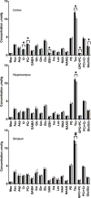
Intrauterine growth restricted (IUGR) infants are at increased risk for neurodevelopmental deficits that suggest the hippocampus and cerebral cortex may be particularly vulnerable. Evaluate regional neurochemical profiles in IUGR and normally grown (NG) 7-day old rat pups using in vivo 1H magnetic resonance (MR) spectroscopy at 9.4 T. IUGR was induced via bilateral uterine artery ligation at gestational day 19 in pregnant Sprague-Dawley dams. MR spectra were obtained from the cerebral cortex, hippocampus and striatum at P7 in IUGR (N = 12) and NG (N = 13) rats. In the cortex, IUGR resulted in lower concentrations of phosphocreatine, glutathione, taurine, total choline, total creatine (P < 0.01) and [glutamate]/[glutamine] ratio (P < 0.05). Lower taurine concentrations were observed in the hippocampus (P < 0.01) and striatum (P < 0.05). IUGR differentially affects the neurochemical profile of the P7 rat brain regions. Persistent neurochemical changes may lead to cortex-based long-term neurodevelopmental deficits in human IUGR infants.
Keywords: Brain; IUGR; Magnetic resonance spectroscopy; Metabolism.
Figures


References
-
- Lin CH, Gelardi NL, Cha CJ, Oh W. Cerebral metabolic response to hypoglycemia in severe intrauterine growth-retarded rat pups. Early Hum Dev. 1998;52:1–11. - PubMed
-
- Rosenberg A. The IUGR newborn. Semin Perinatol. 2008;32:219–224. - PubMed
-
- Padilla N, et al. Differential vulnerability of gray matter and white matter to intrauterine growth restriction in preterm infants at 12 months corrected age. Brain Res. 2014;1545:1–11. - PubMed
-
- Baschat AA. Neurodevelopment following fetal growth restriction and its relationship with antepartum parameters of placental dysfunction. Ultrasound Obstet Gynecol. 2011;37:501–514. - PubMed
-
- Leitner Y, et al. Neurodevelopmental outcome of children with intrauterine growth retardation: a longitudinal, 10-year prospective study. J Child Neurol. 2007;22:580–587. - PubMed
MeSH terms
Grants and funding
LinkOut - more resources
Full Text Sources
Other Literature Sources

