Review
. 2016 Feb;44(1):1-10.
doi: 10.1007/s15010-015-0780-z. Epub 2015 May 14.

Chest ultrasonography in patients with HIV: a case series and review of the literature

Affiliations
Review

Chest ultrasonography in patients with HIV: a case series and review of the literature

Charlotte C Heuvelings et al. Infection. 2016 Feb.

Abstract

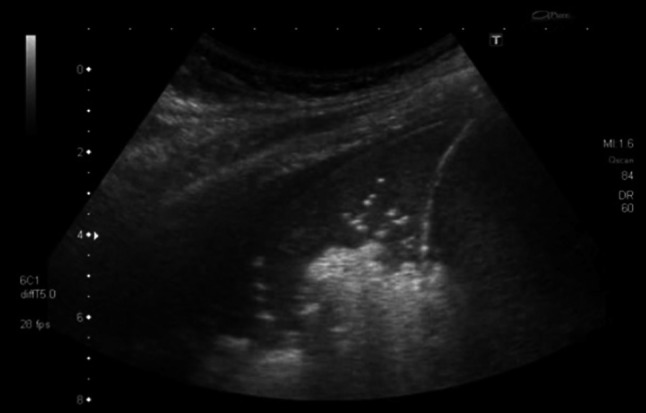
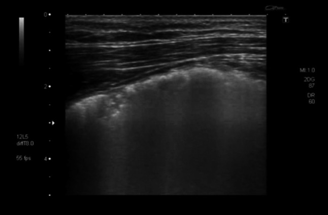
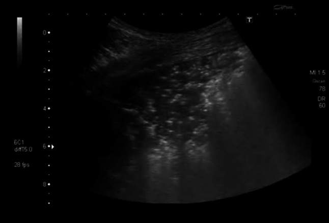
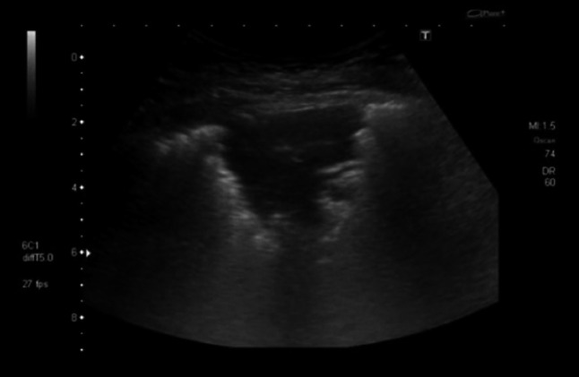
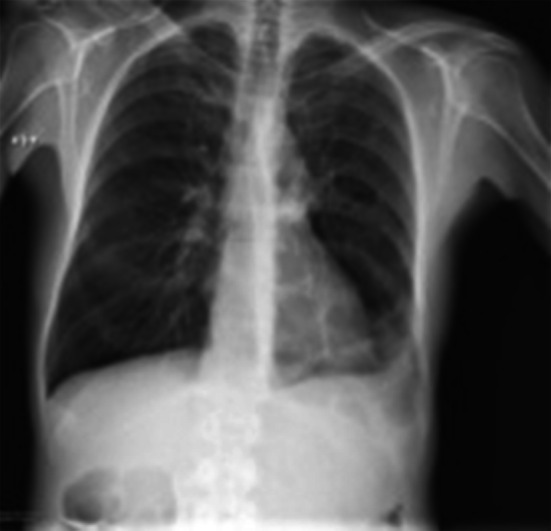
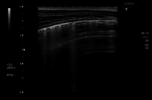
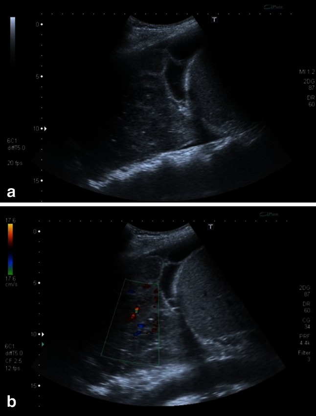
Introduction: Pulmonary disease is common in HIV-infected patients. Diagnostic means, however, are often scarce in areas where most HIV patients are living. Chest ultrasonography has recently evolved as a highly sensitive and specific imaging tool for diagnosing chest conditions such as pneumothorax, pneumonia and pulmonary edema in critically ill patients. This article addresses the issue of imaging and differentiating common pulmonary conditions in HIV-infected patients by chest ultrasonography.

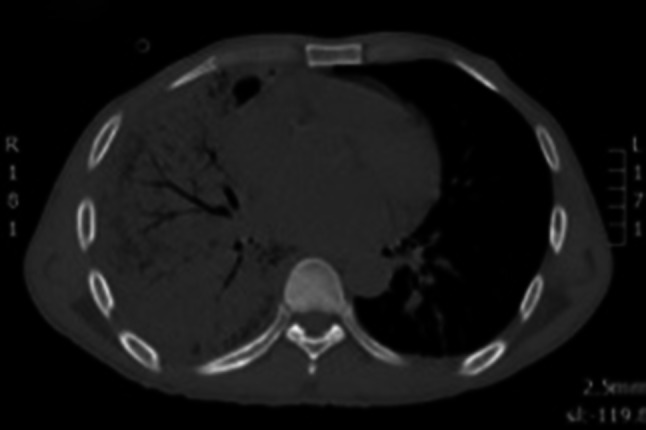
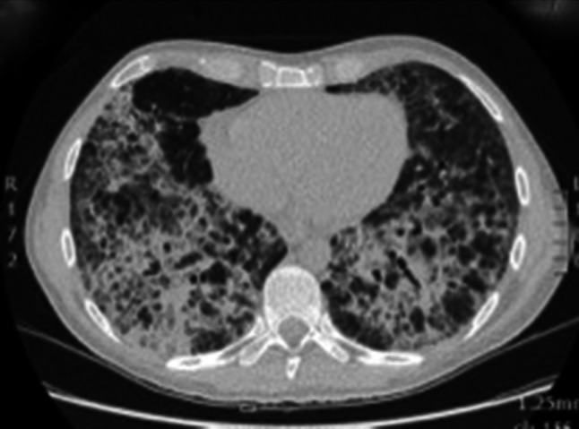
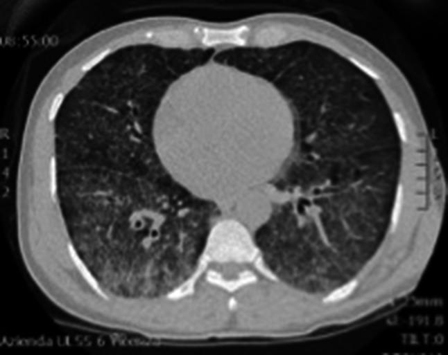
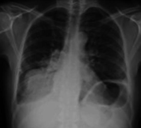
Methods: We report chest ultrasound features of five different common pulmonary diseases in HIV-infected patients (bacterial pneumonia, Pneumocystis jirovecii pneumonia, tuberculosis, cytomegalovirus pneumonia and non-Hodgkin lymphoma) and review the respective literature.

Conclusions: We observed characteristic ultrasound patterns especially in Pneumocystis jirovecii pneumonia and pulmonary lymphoma. Further exploration of chest ultrasonography in HIV-infected patients appears promising and may translate into new diagnostic approaches for pulmonary conditions in patients living with HIV.

Keywords: Chest ultrasonography; Diagnostic imaging; HIV; Infection; Lung disease; Pulmonary.


Figures

Fig. 1
Fig. 1
Lung CT scan showing right basal consolidation and air bronchograms
Fig. 2
Fig. 2
Transthoracic ultrasonography showing a hypoechoic consolidation with string-like echogenic reflexes due to remaining air in the bronchial system (sonographic air bronchogram). Additionally, a small pleural effusion is visible
Fig. 3
Fig. 3
Transthoracic ultrasonography of the pleura shows an interstitial pattern with multiple B-lines. Additionally, small subpleural consolidations are present
Fig. 4
Fig. 4
Chest CT after development of respiratory distress showing coarse interstitial thickening with cyst formation and areas of unaffected parenchyma suggestive of pneumocystis pneumonia, which was later microbiologically confirmed in BAL
Fig. 5
Fig. 5
Transthoracic ultrasonography showing large consolidated areas with multiple bright artifacts suggesting gas inclusions. These represent both air bronchograms and cystic areas of the lung
Fig. 6
Fig. 6
Transthoracic ultrasonography showing a subpleural hypoechoic consolidation. Due to the shredded, fractal boundary between the consolidation and the underlying aerated lung this is called “shred sign”
Fig. 7
Fig. 7
Chest radiograph of a febrile HIV-infected male (CD4 count 173 cells/μl) suggesting a left sided basal pneumonia
Fig. 8
Fig. 8
Transthoracic ultrasonography of the pleura. Multiple B-lines are visible suggesting an interstitial pattern of lung injury; as these are not penetrating the complete image, nor do they extinguish the A-lines completely which are visible towards the right side of the screen, these should be referred to as “lung rockets”
Fig. 9
Fig. 9
Chest CT scan showing ground-glass appearance and interstitial thickening due to interstitial pneumonia caused by the underlying generalized CMV infection
Fig. 10
Fig. 10
Chest radiograph with consolidation adjacent to the right hemi-diaphragm
Fig. 11
Fig. 11
a Sonographic image of a right-sided consolidation. The consolidation shows areas of lower echogenicity suggesting lymphoma infiltration. Additionally a pleural effusion with fibrin strands is present. b Using color-Doppler the hypoechoic areas surround the vasculature (see Online Resources 6 and 7)

References

    1. Moore CL, Copel JA. Point-of-care ultrasonography. N Engl J Med. 2011;364:749–757. doi: 10.1056/NEJMra0909487. - DOI - PubMed
    1. Ding W, Shen Y, Yang J, He X, Zhang M. Diagnosis of pneumothorax by radiography and ultrasonography: a meta-analysis. Chest. 2011;140:859–866. doi: 10.1378/chest.10-2946. - DOI - PubMed
    1. Lichtenstein DA, Meziere G, Lascols N, Biderman P, Courret JP, Gepner A, Goldstein I, Tenoudji-Cohen M. Ultrasound diagnosis of occult pneumothorax. Crit Care Med. 2005;33:1231–1238. doi: 10.1097/01.CCM.0000164542.86954.B4. - DOI - PubMed
    1. Knudtson JL, Dort JM, Helmer SD, Smith RS. Surgeon-performed ultrasound for pneumothorax in the trauma suite. J Trauma. 2004;56:527–530. doi: 10.1097/01.TA.0000114529.99353.22. - DOI - PubMed
    1. Copetti R, Cattarossi L. Ultrasound diagnosis of pneumonia in children. Radiol Med (Torino) 2008;113:190–198. doi: 10.1007/s11547-008-0247-8. - DOI - PubMed

Publication types

MeSH terms

LinkOut - more resources