. 2015 May 13;35(19):7365-73.
doi: 10.1523/JNEUROSCI.4793-14.2015.

Neural mechanisms of placebo anxiolysis

Affiliations

Neural mechanisms of placebo anxiolysis

Benjamin Meyer et al. J Neurosci. .

Abstract

The beneficial effects of placebo treatments on fear and anxiety (placebo anxiolysis) are well known from clinical practice, and there is strong evidence indicating a contribution of treatment expectations to the efficacy of anxiolytic drugs. Although clinically highly relevant, the neural mechanisms underlying placebo anxiolysis are poorly understood. In two studies in humans, we tested whether the administration of an inactive treatment along with verbal suggestions of anxiolysis can attenuate experimentally induced states of phasic fear and/or sustained anxiety. Phasic fear is the response to a well defined threat and includes attentional focusing on the source of threat and concomitant phasic increases of autonomic arousal, whereas in sustained states of anxiety potential and unclear danger requires vigilant scanning of the environment and elevated tonic arousal levels. Our placebo manipulation consistently reduced vigilance measured in terms of undifferentiated reactivity to salient cues (indexed by subjective ratings, skin conductance responses and EEG event-related potentials) and tonic arousal [indexed by cue-unrelated skin conductance levels and enhanced EEG alpha (8-12 Hz) activity], indicating a downregulation of sustained anxiety rather than phasic fear. We also observed a placebo-dependent sustained increase of frontal midline EEG theta (4-7 Hz) power and frontoposterior theta coupling, suggesting the recruitment of frontally based cognitive control functions. Our results thus support the crucial role of treatment expectations in placebo anxiolysis and provide insight into the underlying neural mechanisms.

Keywords: EEG; anxiety; event-related potentials; frontal midline theta; placebo effect.


Figures

Figure 1.
Figure 1.
Study design. The study used a two by two factorial design with factors threat (NT, T) and placebo (NP, P). An experiment comprised six runs in which participants were under the verbally induced illusion to receive an anxiolytic pharmacological treatment (P runs) and six runs without placebo (NP runs). Each run began with the intake of either of one nasal sprays labeled “L” (in P runs) and “N” (in NP runs). Within each run, there were 11–12 trials of T (red square) or NT (green square), each of 5 s duration. During T trials, participants knew they receive a painful electric shock with a probability of 25%. We reasoned that T trials compared with NT trials induce a phasic fear reaction, whereas the temporally unpredictable occurrence of T trials within runs created an uncertain, threatening context that induces sustained anxiety. At the end of each run, participants were asked to rate their fear for the red and green squares. Skin conductance (in Studies 1 and 2) and EEG (in Study 2) were measured concurrently.
Figure 2.
Figure 2.
Example of a decomposed skin conductance time course. SC time course and underlying tonic component (SCtonic) are represented by black and gray lines, respectively. The phasic component (SC phasic) can be obtained by subtracting SCtonic (gray line) from SC (black line). Dashed lines indicate trial onsets of T (red squares) and NT trials (green squares). Painful electric stimuli were applied in Trials 8 and 12.
Figure 3.
Figure 3.
Behavioral results (Studies 1 and 2). In both studies, fear ratings (A, D) and SCRs (B, E) to the cues revealed main effects of placebo (P-NP), indicating a cue-unspecific placebo effect. Tonic SCLs measured throughout experimental runs (C, F) showed reduced arousal under placebo (P-NP); *p < 0.05; **p < 0.01; ***p < 0.001. Error bars indicate SEM.
Figure 4.
Figure 4.
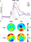
ERPs (Study 2). Average activation time courses of electrode P4 time-locked to cue onset (0 s). Significant main effects of threat (T-NT, red-blue curves) and placebo (P-NP, light-dark curves) were found for the P300 and for the LPP components (A). Topographic voltage difference maps for the threat and placebo main effects (T-NT: voltage increases, red; P-NP: voltage reductions, blue) in P300 and LPP (B). White dots indicate electrodes of significant clusters (p < 0.05).
Figure 5.
Figure 5.
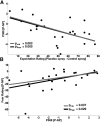
Brain-behavior correlations (Study 2). The reduction of P300 amplitudes under placebo in electrode P4 was predicted by pre-experimental treatment expectations (A) and also correlated with the placebo main effects in fear ratings across participants (B). pPear, p Value of Pearson's correlation; prob, p value of robust correlation.
Figure 6.
Figure 6.
ITI alpha activity (Study 2). ITI alpha activity was increased in placebo relative to no placebo runs (P-NP) for the total sample. Only HRs but not LRs showed increased alpha activity in placebo runs. A widespread frontal cluster revealed significant differences between HRs and LRs (HR-LR). White dots indicate electrodes of significant clusters (p < 0.05).
Figure 7.
Figure 7.
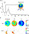
ITI theta activity (Study 2). Illustration of the ITI power spectrum collapsed across P and NP runs of electrode Fz and the average of all electrodes (Av). Overall power in the theta band was elevated in frontal midline channels including electrode Fz (A). FMT power was increased during ITIs in placebo relative to no-placebo runs (P-NP) for the total sample (left column). FPTC was also enhanced in placebo runs. Middle and right columns: HR and LR placebo responders. Significant coupling between cluster electrodes and electrode Fz is indicated by dashed lines. Noncluster electrodes are masked in FPTC results (B).

Similar articles

Cited by

References

    1. Aftanas LI, Golocheikine SA. Human anterior and frontal midline theta and lower alpha reflect emotionally positive state and internalized attention: high-resolution EEG investigation of meditation. Neurosci Lett. 2001;310:57–60. doi: 10.1016/S0304-3940(01)02094-8. - DOI - PubMed
    1. Amanzio M, Benedetti F, Porro CA, Palermo S, Cauda F. Activation likelihood estimation meta-analysis of brain correlates of placebo analgesia in human experimental pain. Hum Brain Mapp. 2013;34:738–752. doi: 10.1002/hbm.21471. - DOI - PMC - PubMed
    1. Anguera JA, Boccanfuso J, Rintoul JL, Al-Hashimi O, Faraji F, Janowich J, Kong E, Larraburo Y, Rolle C, Johnston E, Gazzaley A. Video game training enhances cognitive control in older adults. Nature. 2013;501:97–101. doi: 10.1038/nature12486. - DOI - PMC - PubMed
    1. Baas JM, Kenemans JL, Böcker KB, Verbaten MN. Threat-induced cortical processing and startle potentiation. Neuroreport. 2002;13:133–137. doi: 10.1097/00001756-200201210-00031. - DOI - PubMed
    1. Benedek M, Kaernbach C. A continuous measure of phasic electrodermal activity. J Neurosci Methods. 2010;190:80–91. doi: 10.1016/j.jneumeth.2010.04.028. - DOI - PMC - PubMed

Publication types

MeSH terms