. 2015 May 13;35(19):7487-502.
doi: 10.1523/JNEUROSCI.4454-14.2015.

Synapsin determines memory strength after punishment- and relief-learning

Affiliations

Synapsin determines memory strength after punishment- and relief-learning

Thomas Niewalda et al. J Neurosci. .

Abstract

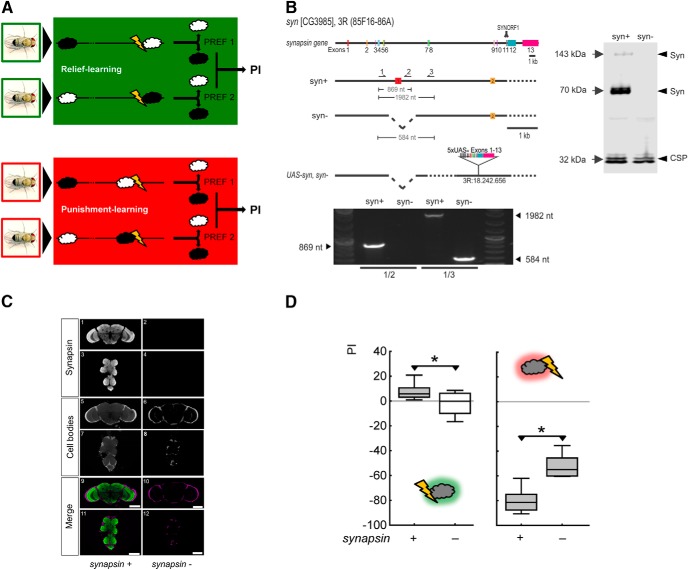
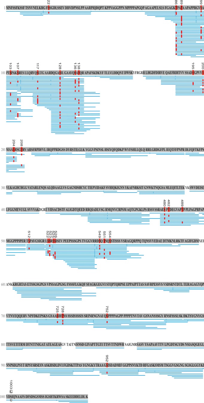
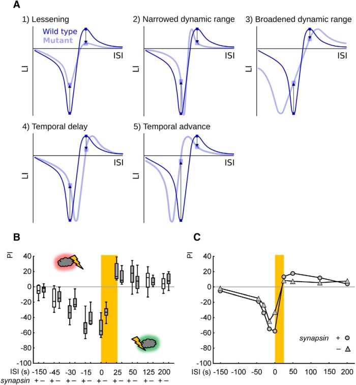
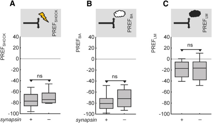
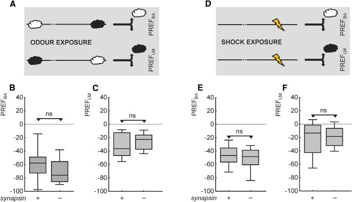
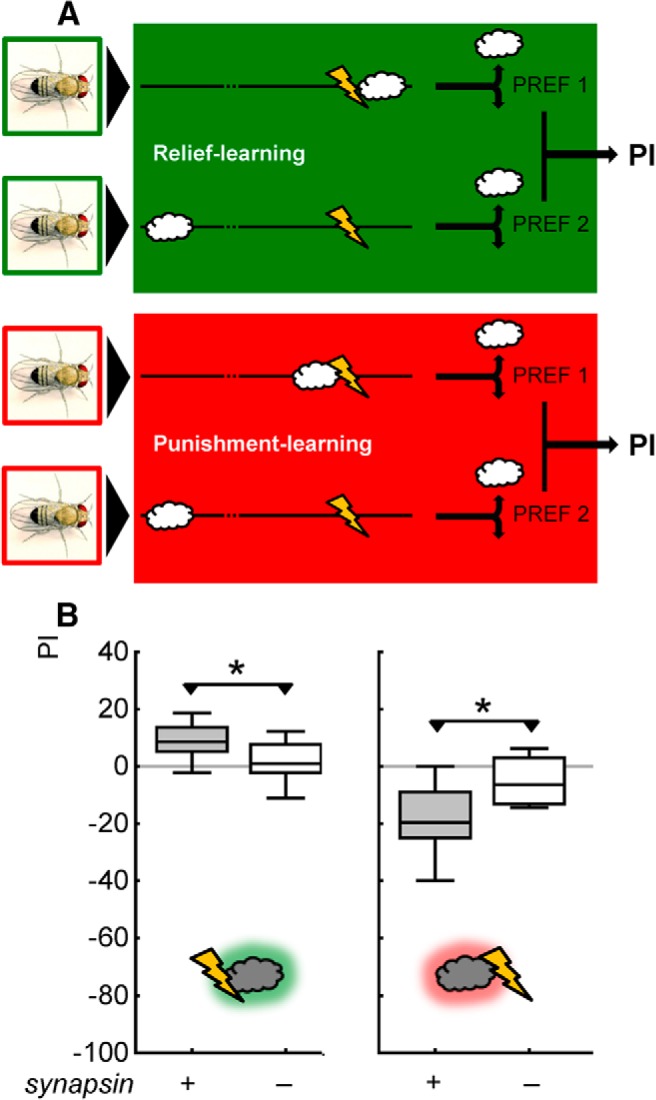
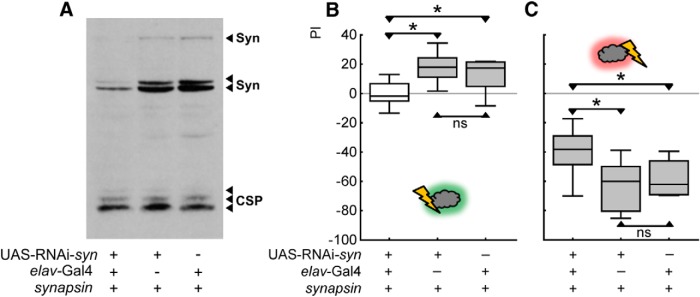
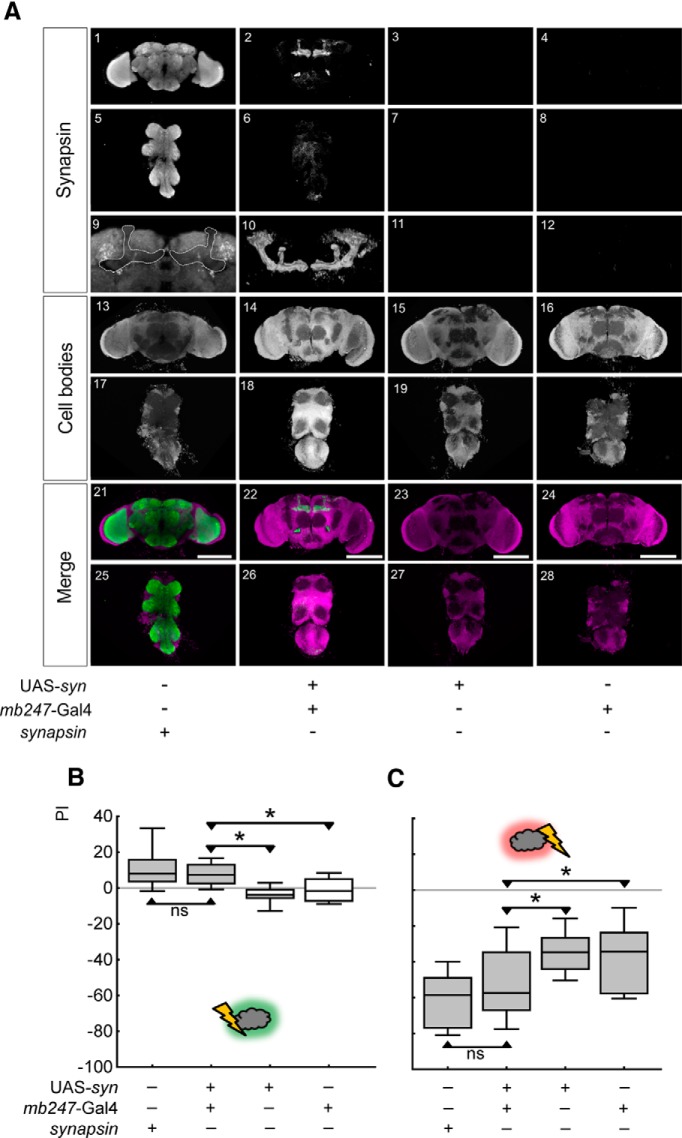
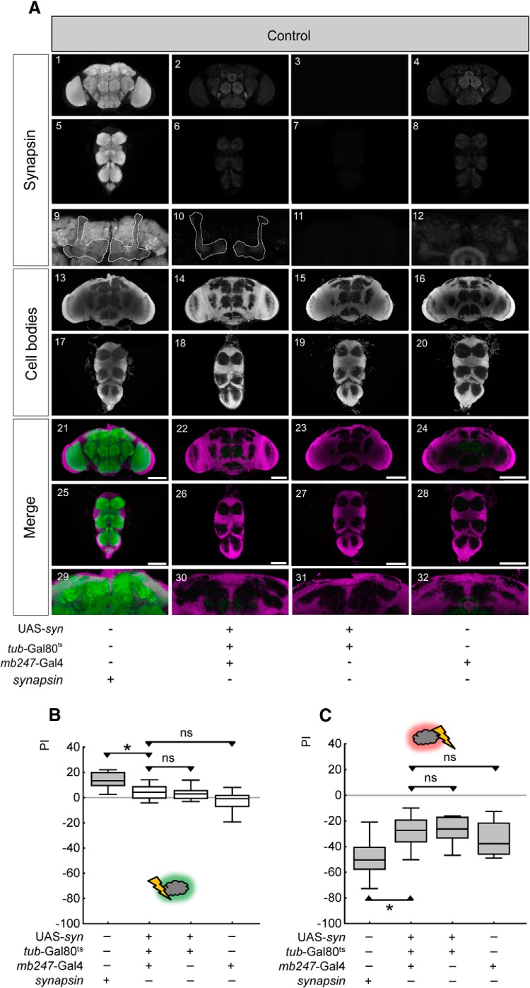
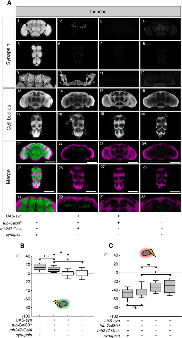
Adverse life events can induce two kinds of memory with opposite valence, dependent on timing: "negative" memories for stimuli preceding them and "positive" memories for stimuli experienced at the moment of "relief." Such punishment memory and relief memory are found in insects, rats, and man. For example, fruit flies (Drosophila melanogaster) avoid an odor after odor-shock training ("forward conditioning" of the odor), whereas after shock-odor training ("backward conditioning" of the odor) they approach it. Do these timing-dependent associative processes share molecular determinants? We focus on the role of Synapsin, a conserved presynaptic phosphoprotein regulating the balance between the reserve pool and the readily releasable pool of synaptic vesicles. We find that a lack of Synapsin leaves task-relevant sensory and motor faculties unaffected. In contrast, both punishment memory and relief memory scores are reduced. These defects reflect a true lessening of associative memory strength, as distortions in nonassociative processing (e.g., susceptibility to handling, adaptation, habituation, sensitization), discrimination ability, and changes in the time course of coincidence detection can be ruled out as alternative explanations. Reductions in punishment- and relief-memory strength are also observed upon an RNAi-mediated knock-down of Synapsin, and are rescued both by acutely restoring Synapsin and by locally restoring it in the mushroom bodies of mutant flies. Thus, both punishment memory and relief memory require the Synapsin protein and in this sense share genetic and molecular determinants. We note that corresponding molecular commonalities between punishment memory and relief memory in humans would constrain pharmacological attempts to selectively interfere with excessive associative punishment memories, e.g., after traumatic experiences.

Keywords: Drosophila; Synapsin; memory; pain; punishment; relief.


Figures

Figure 1.
Figure 1.
Mutant flies lacking Synapsin are impaired in both relief memory and punishment memory. A, Schematic of the experimental paradigm for relief learning and punishment learning. For relief learning, an odor is presented upon the cessation of shock, whereas for punishment learning an odor is presented before the onset of shock. In both cases, a control odor is presented temporally far removed from shock. During the test, the flies can choose between the relief-trained versus the control odor, and between the punishment-trained versus the control odor, respectively. Swapping the chemical identity of the odors in reciprocally trained flies allows a PI to be calculated from the difference in preference between these reciprocally trained flies. The PI thus measures associative memory and runs no risk of confounding differences in innate preference for either odor, or in nonassociative memory components. For further details see Materials and Methods. B, Upper left, Genomic organization of the Drosophila synapsin gene. Syn contains 13 exons and spans a 13.7 kb genomic region in the 3R (85F16–86A) cytological interval [flybase (www.flybase.org); the second of the 14 exons previously suggested on the basis of syn-cDNA sequences (Klagges et al., 1996) apparently represents a splice artifact (B. Klagges and E. Buchner, personal communication)]. Accordingly, the coding region for the epitope LFGGMEVCGL that is recognized by the monoclonal antibody SYNORF1 is encoded by exon 11. The syn97CS strain (labeled synapsin for simplicity) carries a 1.4 kb deletion spanning parts of the regulatory region and the first exon of the synapsin gene. The arrows indicate the binding sites for the PCR primers upstream (1), within (2), and downstream (3) of the deletion. The effector strain UAS-syn, synapsin contains the syn-cDNA plus 156 base pairs upstream (indicated by * between UAS and Exons 1–13). Lower left, In a single-fly PCR approach, primer combination 1/2 yields an 869 nt product in synapsin+ wild-type but not in synapsin mutants, whereas primer pair 1/3 yields a 1982 nt product in synapsin+ and a 584 nt product in synapsin. Right, Western blot from material obtained from three adult fly heads stained for Synapsin and for CSP as a loading control. The single band at ∼143 kDa and the double band at ∼70 kDa, where Synapsin isoforms are expected (Klagges et al., 1996), are found in synapsin+ but not in synapsin flies. C, Synapsin immunoreactivity is absent in whole-mount preparations of synapsin mutant flies. In the bottom row (Merge), anti-Synapsin staining is shown in green, and cell-body counterstaining with propidium iodide in magenta, displayed as frontal optical sections (0.9 μm) of synapsin+ (left column) and synapsin (right column) brain and thoracic nervous system. Scale bar, 100 μm. D, Left, Shows that relief memory is intact in synapsin+ wild-type flies, but is abolished in mutant flies lacking Synapsin (synapsin). Right, Shows that punishment memory is impaired, but is not abolished, in synapsin mutant flies. *p < 0.05 for the between-genotype comparison within an experiment; a gray shading of the boxes indicates p < 0.05/2 in comparisons of either genotype to chance levels (zero) within one experiment. PI, indicating the difference in preference between reciprocally trained flies, and thus learned approach (positive scores) and learned avoidance (negative scores), respectively. The middle line of the box plots represents the median, the box boundaries the 25 and 75% quartiles, and the whiskers the 10 and 90% quantiles, respectively.
Figure 2.
Figure 2.
Phosphorylation sites of Synapsin and abundance of edited and nonedited Synapsin. LC-MS/MS analysis of experimentally naive, wild-type fly brains to map phosphorylated sites across the Synapsin protein (see also Table 1). Eighty-five LC-MS/MS runs were performed, consisting of a combination of 17 biological and five technical replicates each (coverage of the longest Synapsin protein isoform of 97%). This identified 28 phosphorylated sites of Synapsin: one at tyrosine, 10 at threonine, and 17 at serine. Twenty-three phosphorylated sites were identified for the first time, while five had been reported before (Zhai et al., 2008; Nuwal et al., 2011). Of the seven phosphorylated sites both reported by Nuwal et al. (2011) and covered by the present data, we can confirm three, while we found four of these sites to be nonphosphorylated. Both the edited and the nonedited forms of the Synapsin protein were found. That is, the pre-mRNA of the synapsin transcript is modified from the N-terminal motif RRFS (nonedited) to RGFS (edited) such that the PKA consensus motif RRFS is compromised (Diegelmann et al., 2006). In n = 85 LC-MS/MS runs the nonedited protein motif (RRFS) was found 54 times (p = 1.71E-07) and the edited protein motif (RGFS) was found 22 times (p = 9.44E-13). A phosphorylation at the motifs' serine was reliably detected only once (sic) for the edited, but not at all for the nonedited protein motif (Table 1; S22/S6). The workflow optimized sensitivity for proteome and phosphorylation site analysis of sample amounts corresponding to only a single brain. Therefore, a separation of isoforms before MS was not warranted, such that discrimination between isoforms is not possible. Given a 97% coverage, however, it is possible to ascertain the longest isoform (isoform D, E2QCY9_DROME; www.uniprot.org); this D isoform emerges from transcription starting at the first start codon and read through at the first stop codon (Klagges et al., 1996; Jungreis et al., 2011). A shorter isoform based on transcription from the second start codon and thus lacking 16 aa at the N terminus (Q24546_DROME; www.uniprot.org) was confirmed previously (Nuwal et al., 2011). Blue bars below the sequence indicate peptide-spectra matches (PSMs) identified by LC-MS/MS and the PEAKS de novo sequencing algorithm. The red “P” boxes indicate phosphorylation (p < 0.005). Note the S1003 → G mutation (white “G” box).
Figure 3.
Figure 3.
Memory strength is lessened, rather than features of coincidence detection modulated, in mutant flies lacking Synapsin. A, When using only one ISI for relief learning and one ISI for punishment learning, less strong scores in both relief memory and punishment memory could be a result of a lessening in strength of the associative memory (A1), a narrowed dynamic range of associative coincidence detection (A2), a broadened dynamic range (A3), a temporal delay (A4) or a temporal advance (A5), if the ISIs happened to be chosen as indicated by the arrows. We therefore decided to compare the full ISI function between synapsin+ and synapsin mutant flies. B, Associative performance indices of wild-type synapsin+ (respective left plots) and the mutant synapsin flies (respective right plots) for the indicated ISIs. For statistics, see body text. A gray shading of the boxes indicates p < 0.05/10 and p < 0.05/8, respectively, in comparisons to chance levels (zero). Other details as in Figure 1. C, The median PIs from B are plotted across the ISIs. The consistent lessening of scores throughout the ISI function resembles scenario A1, suggesting a lessening of associative memory in the mutant synapsin flies.
Figure 4.
Figure 4.
Behavior toward the to-be-associated stimuli is normal in experimentally naive mutant flies lacking Synapsin. Avoidance of the shock (A) and of the odors (B: BA; C: LM) is not different between experimentally naive flies of the two genotypes. ns: p > 0.05. Other details as in Figure 1.
Figure 5.
Figure 5.
Olfactory behavior is normal in mutant flies lacking Synapsin also after training-like stimulus exposure. Genotypes do not differ in olfactory behavior after either odor exposure (A; B: BA, C: LM) or shock exposure (D; E: BA, F: LM). Other details as in Figure 4.
Figure 6.
Figure 6.
Mutants lacking Synapsin are defective also in nondiscriminatory relief- and punishment-learning tasks. A, Schematic of the one-odor versions of the relief- and punishment-learning tasks. The procedure is as in the two-odor version of the paradigm (Fig. 1A; see Materials and Methods), except that one odor is omitted. That is, for one-odor relief learning, the odor (BA) is presented upon the cessation of shock, while for punishment learning the odor is presented before the onset of shock. In both cases, a second experimental group receives unpaired presentations of odor and punishment. The difference in odor preference between paired- and unpaired-trained groups indicates associative memory, and is quantified as PI. B, Also in nondiscriminatory, one-odor versions of the paradigm, relief memory and punishment memory are strongly impaired in mutant flies lacking Synapsin (synapsin). Other details as in Figure 1D.
Figure 7.
Figure 7.
RNAi-mediated knockdown of Synapsin impairs both relief memory and punishment memory. A, Western blot of material obtained from three heads stained for Synapsin and for CSP as a loading control. The blot is loaded with double heterozygous elav-Gal4;; UAS-RNAi-syn flies to the left (knockdown), UAS-RNAi-syn heterozygous flies in the middle (effector control), and elav-Gal4 heterozygote flies to the right (driver control). In the knock-down flies, a reduction of all Synapsin isoforms is apparent. B, Relief memory is abolished in knock-down flies compared with controls. C, Punishment memory is reduced in knock-down flies compared with controls. *p < 0.05/3 and ns: p > 0.05/3 are used for pairwise comparisons. Gray shading of the boxes indicates significance from chance (zero) at p < 0.05/3.
Figure 8.
Figure 8.
Locally restoring Synapsin restores relief memory and punishment memory. A, Expression pattern of Synapsin in flies of the indicated genotypes. In the bottom row (Merge), anti-Synapsin staining in brains and thoracic nervous systems is shown in green, and cell-body counterstaining with propidium iodide is shown in magenta, from 0.9 μm frontal optical sections of the indicated genotypes. In A9–12, the mushroom body regions from A1–4 are shown at higher magnification. In A9 the mushroom bodies are indicated by the stippled line. In A10 the expression of Synapsin using the mb247-Gal4 driver is shown as a 3D display. Scale bars: 100 μm. B, Relief memory of synapsin mutant flies is fully restored upon rescue expression of Synapsin using the mb247-Gal4 driver. C, Punishment memory, too, is fully restored upon locally expressing Synapsin. *p < 0.05/3 and ns: p > 0.05/3 are used for pairwise comparisons. Gray shading of the boxes indicates significance from chance (zero) at p < 0.05/4.
Figure 9.
Figure 9.
Relief memory and punishment memory remain impaired in control conditions without acute and local restoration of Synapsin. A, Expression pattern of Synapsin in uninduced control flies of the indicated genotypes. In the bottom rows (Merge), anti-Synapsin staining in brains and thoracic nervous systems is shown in green, while cell-body counterstaining with propidium iodide is shown in magenta. The mushroom body region of A1–4 is shown at higher magnification in A9–12. The stippled line in A9 and A10 indicates the mushroom body neuropil. The mushroom body region of A21–24 is shown at higher magnification in A29–32. Note the absence of anti-Synapsin staining in A10 and A30. Scale bars: 100 μm. B, C, In uninduced control conditions, relief memory (B) and punishment memory (C) remain abolished in the experimental genotype. *p < 0.05/3 and ns: p > 0.05/3 are used for pairwise comparisons. Gray shading of the boxes indicates significance from chance (zero) at p < 0.05/4.
Figure 10.
Figure 10.
Acutely and locally inducing Synapsin expression restores relief memory and punishment memory. A, Expression pattern of Synapsin in induced rescue flies of the indicated genotypes. In the bottom row (Merge), anti-Synapsin staining in brains and thoracic nervous systems is shown in green, while cell-body counterstaining with propidium iodide is shown in magenta. The mushroom body region of A1–4 is shown at higher magnification in A9–12. The stippled line in A9 indicates the mushroom body neuropil. In A10 the expression of Synapsin induced by the mb247-Gal4 driver is shown as a 3D display. The mushroom body region of A21–24 is shown at higher magnification in A29–32. Scale bars: 100 μm. B, Relief memory of synapsin mutant flies is restored upon acutely induced local expression of Synapsin using the mb247-Gal4 driver in combination with Gal80ts. C, Punishment memory, too, is fully restored upon acutely and locally expressing Synapsin. *p < 0.05/3 and ns: p > 0.05/3 are used for pairwise comparisons. Gray shading of the boxes indicates significance from chance (zero) at p < 0.05/4.

Similar articles

Cited by

References

    1. Andreatta M, Mühlberger A, Yarali A, Gerber B, Pauli P. A rift between implicit and explicit conditioned valence in human pain relief learning. Proc Biol Sci. 2010;277:2411–2416. doi: 10.1098/rspb.2010.0103. - DOI - PMC - PubMed
    1. Andreatta M, Fendt M, Mühlberger A, Wieser MJ, Imobersteg S, Yarali A, Gerber B, Pauli P. Onset and offset of aversive events establish distinct memories requiring fear and reward networks. Learn Mem. 2012;19:518–526. doi: 10.1101/lm.026864.112. - DOI - PubMed
    1. Arnold C, Reisch N, Leibold C, Becker S, Prüfert K, Sautter K, Palm D, Jatzke S, Buchner S, Buchner E. Structure–function analysis of the cysteine string protein in Drosophila: cysteine string, linker and C terminus. J Exp Biol. 2004;207:1323–1334. doi: 10.1242/jeb.00898. - DOI - PubMed
    1. Aso Y, Grübel K, Busch S, Friedrich AB, Siwanowicz I, Tanimoto H. The mushroom body of adult Drosophila characterized by GAL4 drivers. J Neurogenet. 2009;23:156–172. doi: 10.1080/01677060802471718. - DOI - PubMed
    1. Aso Y, Siwanowicz I, Bräcker L, Ito K, Kitamoto T, Tanimoto H. Specific dopaminergic neurons for the formation of labile aversive memory. Curr Biol. 2010;20:1445–1451. doi: 10.1016/j.cub.2010.06.048. - DOI - PMC - PubMed

Publication types

MeSH terms

LinkOut - more resources