. 2015 Jun 15;194(12):5595-8.
doi: 10.4049/jimmunol.1500623. Epub 2015 May 13.

Cutting edge: Role of osteopontin and integrin αv in T cell-mediated anti-inflammatory responses in endotoxemia

Affiliations

Cutting edge: Role of osteopontin and integrin αv in T cell-mediated anti-inflammatory responses in endotoxemia

Makoto Inoue et al. J Immunol. .

Abstract

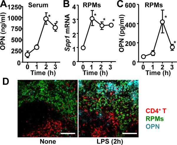
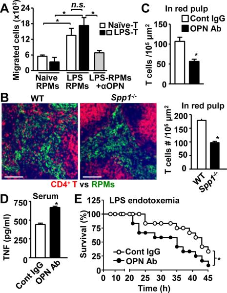
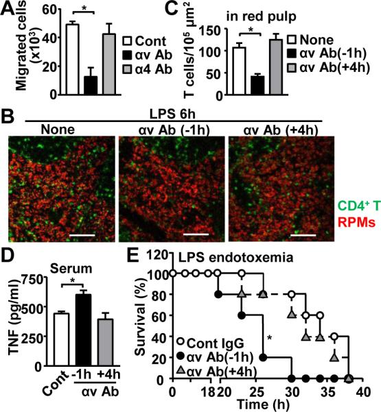
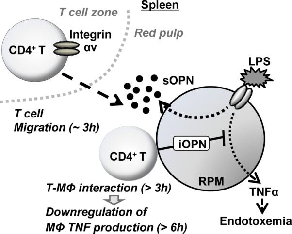
The immune system is equipped with mechanisms that downregulate hyperinflammation to avoid collateral damage. We demonstrated recently that unprimed T cells downregulate macrophage TNF production through direct interaction with macrophages in the spleen during LPS endotoxemia. How T cell migration toward macrophages occurs upon LPS injection is still not clear. In this study, we demonstrate that secreted osteopontin (sOPN) plays a role in the T cell migration to initiate the suppression of hyperinflammation during endotoxemia. Osteopontin levels in splenic macrophages were upregulated 2 h after LPS treatment, whereas T cell migration toward macrophages was observed 3 h after treatment. Neutralization of sOPN and blockade of its receptor, integrin αv, significantly inhibited CD4(+) T cell migration and increased susceptibility to endotoxemia. Our study demonstrates that the sOPN/integrin αv axis, which induces T cell chemotaxis toward macrophages, is critical for suppressing hyperinflammation during the first 3 h of endotoxemia.


Figures

Figure 1
Figure 1. Upregulation of secreted osteopontin (sOPN) in splenic macrophage during LPS endotoxemia
(A) Serum OPN levels at indicated timepoints after LPS i.p. injection (40 mg/kg mouse weight). (B) Spp1 mRNA levels in RPMs isolated at indicated timepoints after LPS i.p. injection. (C) sOPN levels in supernatants of RPM culture. RPMs were isolated at indicated timepoints after LPS i.p. injection, and cultured for 3h in RPMI complete medium before harvesting supernatants. (D) Histological sections of spleens, isolated from naïve mice and LPS-injected mice (2 h after after i.p. injection), were stained to detect OPN (cyan), CD4+ T cells (CD4; red), and RPMs (F4/80; green). All the experiments are representatives from at least 2 similar experiments for each. *; p<0.05.
Figure 2
Figure 2. Requirement of sOPN for T cell migration and control of hyper-inflammation during LPS endotoxemia
(A) Ex vivo migration assay. Splenic T cells and RPMs were isolated from naïve and mice 2 h post LPS injection. OPN neutralizing Ab was added to the bottom chambers in the indicated group. (B) Localization of CD4+ T cells (CD4; red) and RPMs (F4/80; green) in the spleen. Spleen was isolated from wild-type and Spp1−/− mice 3 h after LPS i.p. injection. Representative images (left panels) and results of quantitative analysis (right panel) are shown. Scale bars denote 100 μm. (C) Mice were i.p. treated with OPN Ab 1h prior to LPS injection, and spleens were harvested 3 h after LPS injection. T cell numbers were enumerated in images of the red pulp. Shown are average values of 10 sections/mouse from 3 mice. (D, E) LPS was i.p. injected into mice with (●) or without (○) OPN neutralization Ab i.p. injection 1h prior to LPS injection. n=12. Serum TNFα levels 6 h after injection (D) and survival (E) are shown. *; p<0.05 compared with control mice.
Figure 3
Figure 3. Integrin αv on T cells is critical for T cell migration and resistance to enodotoxemia
(A) Ex vivo T cell migration towards OPN. T cells were pre-treated with blocking Ab of either integrin αv or α4. (B-E) In vivo integrin αv Ab treatment during endotoxemia. Ab was i.p. administered either 1 h before or 4 h after LPS injection. Spleens were harvested at 6 h after LPS injection (B-D). Representative images of spleen (B) and results from T cell enumeration in red plup (C) are shown. Scale bars denote 100 μm. Serum TNFα levels 6 h after LPS injection (D). Mouse survival with (●, ▲) or without (○) integrin αv Ab (E). At least 5 mice/group. *; p<0.05 compared with control mice.
Figure 4
Figure 4. Roles of sOPN and iOPN during LPS endotoxemia
T cells and macrophages are separately localized in different zones in the spleen. OPN expression is upregulated by LPS in RPMs, and sOPN is secreted from LPS-stimulated RPMs in the first 3 h after LPS stimulation. sOPN is detected by integrin αv on the CD4+ T cell surface; and T cells start migrating towards macrophages. T cells then interact with macrophages to stimulate macrophage CD40 signaling pathway, in which intracellular osteopontin (iOPN) is essential for downregulation of macrophage TNF production (9).

References

    1. Kawagoe T, Takeuchi O, Takabatake Y, Kato H, Isaka Y, Tsujimura T, Akira S. TANK is a negative regulator of Toll-like receptor signaling and is critical for the prevention of autoimmune nephritis. Nature immunology. 2009;10:965–972. - PMC - PubMed
    1. Heyninck K, Beyaert R. The cytokine-inducible zinc finger protein A20 inhibits IL-1-induced NF-kappaB activation at the level of TRAF6. FEBS letters. 1999;442:147–150. - PubMed
    1. Heyninck K, De Valck D, Vanden Berghe W, Van Criekinge W, Contreras R, Fiers W, Haegeman G, Beyaert R. The zinc finger protein A20 inhibits TNF-induced NF-kappaB-dependent gene expression by interfering with an RIP- or TRAF2-mediated transactivation signal and directly binds to a novel NF-kappaB-inhibiting protein ABIN. The Journal of cell biology. 1999;145:1471–1482. - PMC - PubMed
    1. Saitoh T, Fujita N, Jang MH, Uematsu S, Yang BG, Satoh T, Omori H, Noda T, Yamamoto N, Komatsu M, Tanaka K, Kawai T, Tsujimura T, Takeuchi O, Yoshimori T, Akira S. Loss of the autophagy protein Atg16L1 enhances endotoxin-induced IL-1beta production. Nature. 2008;456:264–268. - PubMed
    1. Nakahira K, Haspel JA, Rathinam VA, Lee SJ, Dolinay T, Lam HC, Englert JA, Rabinovitch M, Cernadas M, Kim HP, Fitzgerald KA, Ryter SW, Choi AM. Autophagy proteins regulate innate immune responses by inhibiting the release of mitochondrial DNA mediated by the NALP3 inflammasome. Nature immunology. 2011;12:222–230. - PMC - PubMed

Publication types

MeSH terms