. 2015 Aug;89(15):7735-47.
doi: 10.1128/JVI.01017-15. Epub 2015 May 13.

Measles Virus Defective Interfering RNAs Are Generated Frequently and Early in the Absence of C Protein and Can Be Destabilized by Adenosine Deaminase Acting on RNA-1-Like Hypermutations

Affiliations

Measles Virus Defective Interfering RNAs Are Generated Frequently and Early in the Absence of C Protein and Can Be Destabilized by Adenosine Deaminase Acting on RNA-1-Like Hypermutations

Christian K Pfaller et al. J Virol. 2015 Aug.

Abstract

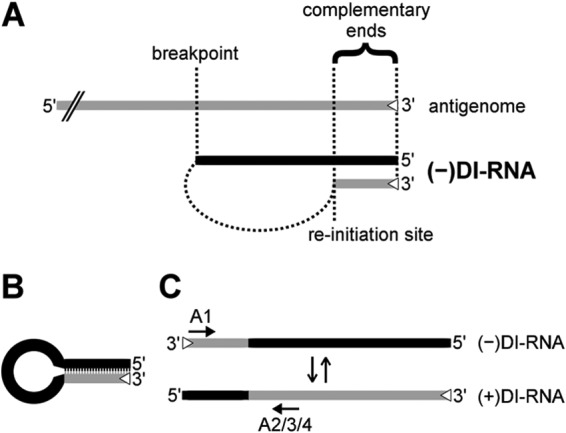
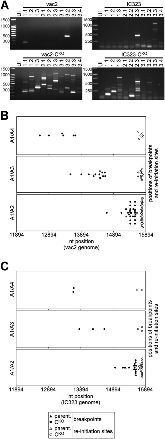
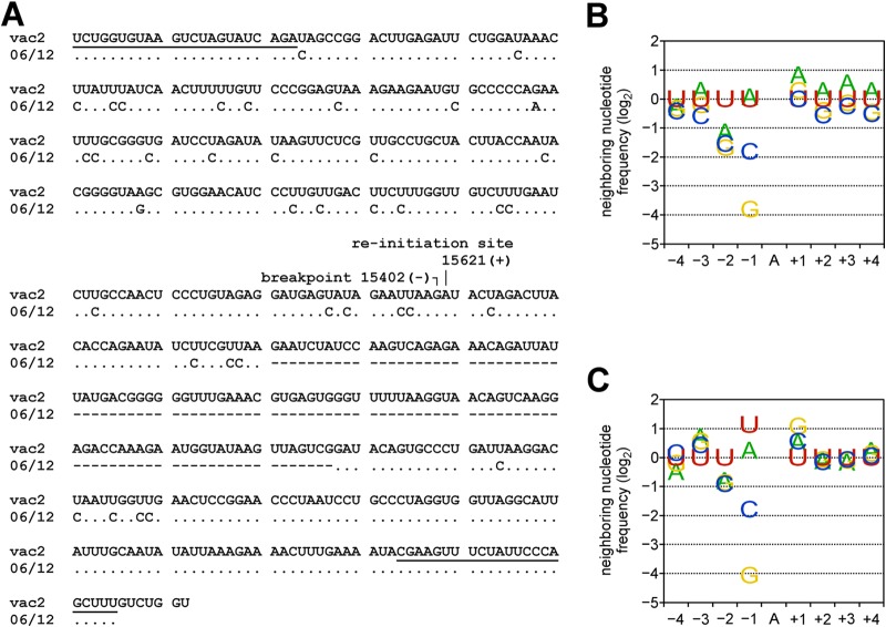
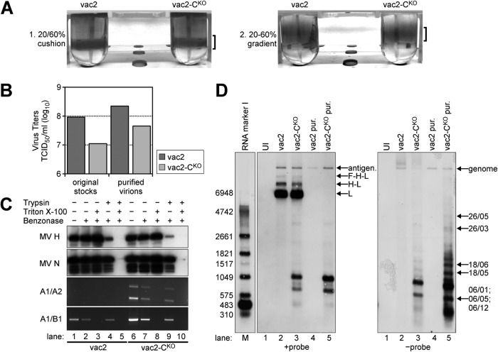
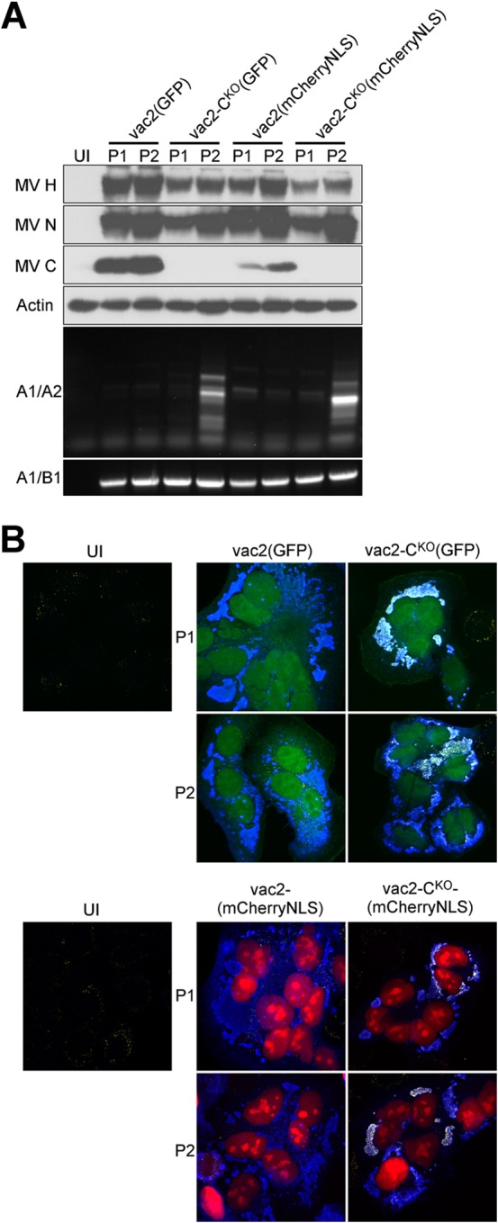
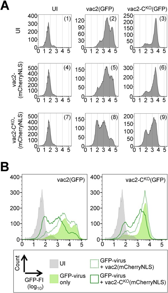
Defective interfering RNAs (DI-RNAs) of the viral genome can form during infections of negative-strand RNA viruses and outgrow full-length viral genomes, thereby modulating the severity and duration of infection. Here we document the frequent de novo generation of copy-back DI-RNAs from independent rescue events both for a vaccine measles virus (vac2) and for a wild-type measles virus (IC323) as early as passage 1 after virus rescue. Moreover, vaccine and wild-type C-protein-deficient (C-protein-knockout [CKO]) measles viruses generated about 10 times more DI-RNAs than parental virus, suggesting that C enhances the processivity of the viral polymerase. We obtained the nucleotide sequences of 65 individual DI-RNAs, identified breakpoints and reinitiation sites, and predicted their structural features. Several DI-RNAs possessed clusters of A-to-G or U-to-C transitions. Sequences flanking these mutation sites were characteristic of those favored by adenosine deaminase acting on RNA-1 (ADAR1), which catalyzes in double-stranded RNA the C-6 deamination of adenosine to produce inosine, which is recognized as guanosine, a process known as A-to-I RNA editing. In individual DI-RNAs the transitions were of the same type and occurred on both sides of the breakpoint. These patterns of mutations suggest that ADAR1 edits unencapsidated DI-RNAs that form double-strand RNA structures. Encapsidated DI-RNAs were incorporated into virus particles, which reduced the infectivity of virus stocks. The CKO phenotype was dominant: DI-RNAs derived from vac2 with a CKO suppressed the replication of vac2, as shown by coinfections of interferon-incompetent lymphatic cells with viruses expressing different fluorescent reporter proteins. In contrast, coinfection with a C-protein-expressing virus did not counteract the suppressive phenotype of DI-RNAs.

Importance: Recombinant measles viruses (MVs) are in clinical trials as cancer therapeutics and as vectored vaccines for HIV-AIDS and other infectious diseases. The efficacy of MV-based vectors depends on their replication proficiency and immune activation capacity. Here we document that copy-back defective interfering RNAs (DI-RNAs) are generated by recombinant vaccine and wild-type MVs immediately after rescue. The MV C protein interferes with DI-RNA generation and may enhance the processivity of the viral polymerase. We frequently detected clusters of A-to-G or U-to-C transitions and noted that sequences flanking individual mutations contain motifs favoring recognition by the adenosine deaminase acting on RNA-1 (ADAR1). The consistent type of transitions on the DI-RNAs indicates that these are direct substrates for editing by ADAR1. The ADAR1-mediated biased hypermutation events are consistent with the protein kinase R (PKR)-ADAR1 balancing model of innate immunity activation. We show by coinfection that the C-defective phenotype is dominant.


Figures

FIG 1
FIG 1
Schematic drawings of 5′ copy-back DI-RNA generation, predicted secondary structure, and replication. (A) 5′ copy-back DI-RNAs are hybrids of truncated genome (black) and antigenome (gray) sequences. During replication, viral polymerase initiates RNA synthesis at the 3′ antigenome promoter (trailer; open triangle) and prematurely terminates synthesis at the breakpoint. The polymerase then reinitiates synthesis on the newly generated strand and finishes replication by recopying the antigenomic 3′ end. The two ends of the DI-RNA are therefore perfectly complementary to each other. (B) Panhandle structure that may be formed through hybridization of the complementary ends of a copy-back DI-RNA in the absence of sufficient N protein. (C) 5′ copy-back DI-RNA replication. The genome DI-RNA [(−)DI-RNA] (top) contains the antigenome promoter in the copy-back sequence (open triangle). Therefore, it can be replicated by the viral polymerase into a complementary antigenome DI-RNA [(+)DI-RNA], which now contains the antigenome promoter at the opposite end. The polarity switch within one DI-RNA molecule allows specific amplification using combinations of primers (primer A1 with either primer A2, A3, or A4; arrows) that would bind in the same direction on a regular antigenome molecule.
FIG 2
FIG 2
Molecular characterization of DI-RNAs generated by vaccine and wild-type MVs. (A) Analysis of DI-RNAs expressed by multiple clones of vaccine MV (vac2) and wild-type MV (IC323) able to express C protein or not (CKO). Plaques were generated in different rescue experiments, amplified, and analyzed by DI-RNA-specific RT-PCR. Uninfected cells (UI) were included as a negative control. The first number above each lane is the experiment number, and the second number is the plaque number. cDNAs were amplified with the A1/A2 primer combination and analyzed by agarose gel electrophoresis. The numbers on the left of each panel indicate the sizes of the marker bands (in base pairs). (B) Nucleotide positions of the breakpoints and reinitiation sites mapped in DI-RNAs derived from vac2 and vac2-CKO (Table 1). The combinations of primers used for amplification are indicated on the left of the three panels. (C) Breakpoint and reinitiation site analysis of IC323 and IC323-CKO viruses (Table 2).
FIG 3
FIG 3
ADAR1-like hypermutations in DI-RNA clones. (A) Alignment of the sequence of DI-RNA clone 06/12 to the vac2 genome (nt 15639 to 15402) and antigenome (nt 15621 to 15887) sequences (GenBank accession number AF266287). The binding sites of the A1 and A2 primers are underlined (top line and bottom two lines, respectively). Identical nucleotides are shown as dots. The breakpoint and reinitiation site nucleotides are indicated. Deleted nucleotides are shown as dashes. (B) Frequency analysis of nucleotides flanking hypermutated sites in DI-RNA clones (all 9 events detected here; Table 3). (C) Frequency of neighboring nucleotides flanking ADAR1 hypermutation sites defined in vitro (21).
FIG 4
FIG 4
Analysis of nucleic acids in vac2- and vac2-CKO-infected cells and in purified vac2 and vac2-CKO particles. (A) Two-step gradient purification of MV particles. Virus bands are indicated by brackets. (B) Titers (numbers of TCID50s per milliliter) of virus stocks prior to and after purification. (C) RNA protection assay with purified virions. Samples were treated with the indicated combinations of trypsin, Triton X-100, and Benzonase; immunoblots were against MV H and N proteins; RT-PCR/PCR analysis was performed for DI-RNAs (A1/A2 primers) and full-length RNA (A1/B1 primers). (D) Northern blot analysis of total RNA from about 3 × 10⁵ uninfected (UI; lane 1) or infected (lanes 2 and 3) Vero cells harvested at 32 h postinfection, as well as RNA extracted from 1 × 10⁶ TCID50s of purified (pur.) and Benzonase-treated vac2 (lane 4) and vac2-CKO (lane 5) particles. The positions of the antigenome and L, H-L, and F-H-L mRNAs (+probe) are indicated on the right side of the left panel. The identities of the genomic RNA and the DI-RNAs (−probe) mapped by PCR amplification are indicated on the right side of the right panel. Lane M, molecular size marker. The numbers on the left side of the left panel indicate the sizes of the marker bands (in bases).
FIG 5
FIG 5
DI-RNA and dsRNA generation by CKO viruses expressing either GFP or mCherryNLS. (A) Western blot and PCR analysis of P1 and P2 of fluorescent reporter-expressing vac2 and vac2-CKO. The antisera used for detection are indicated on the left of the top four panels. Primer pairs used for amplification are indicated on the left of the bottom two panels. UI, uninfected cells. (B) Confocal microscope analysis of HeLa cells infected with fluorescent reporter-expressing vac2 and vac2-CKO. (Top) Autofluorescence of GFP (green); (bottom) autofluorescence of mCherryNLS (red); (all panels) immunofluorescence staining for dsRNA (yellow) and MV N protein (blue). Magnifications, ×40.
FIG 6
FIG 6
Dominance of the CKO phenotype. (A) GFP fluorescence intensities of cells coinfected with viruses indicated on the top and on the left. UI, uninfected cells. Vertical axis, cell counts (linear scale); horizontal axis, GFP fluorescence intensity (FI; log10 scale). (B) Overlay of graphs from panel A. (Left) Summary of coinfections with vac2(GFP); (right) summary of coinfections with vac2-CKO(GFP). Gray area, uninfected cells (panel 1); light green area, single GFP-virus infection (panel 2 or 3); dotted green line, GFP-virus coinfected with vac2(mCherryNLS) (panel 5 or 6); solid green line, GFP-virus coinfected with vac2-CKO(mCherryNLS) (panel 8 or 9).

Similar articles

Cited by

References

    1. Rima BK, Duprex WP. 2009. The measles virus replication cycle. Curr Top Microbiol Immunol 329:77–102. doi:10.1007/978-3-540-70523-9_5. - DOI - PubMed
    1. Lamb RA, Parks GD. 2013. Paramyxoviridae, p 957–995. In Knipe DM, Howley PM, Cohen JI, Griffin DE, Lamb RA, Martin MA, Racaniello VR, Roizman B (ed), Fields virology, 6th ed, vol 1 Lippincott Williams & Wilkins, Philadelphia, PA.
    1. Griffin DE. 2013. Measles virus, p 1042–1069. In Knipe DM, Howley PM, Cohen JI, Griffin DE, Lamb RA, Martin MA, Racaniello VR, Roizman B (ed), Fields virology, 6th ed, vol 1 Lippincott Williams & Wilkins, Philadelphia, PA.
    1. Longhi S. 2009. Nucleocapsid structure and function. Curr Top Microbiol Immunol 329:103–128. doi:10.1007/978-3-540-70523-9_6. - DOI - PubMed
    1. Schrag SJ, Rota PA, Bellini WJ. 1999. Spontaneous mutation rate of measles virus: direct estimation based on mutations conferring monoclonal antibody resistance. J Virol 73:51–54. - PMC - PubMed

Publication types

MeSH terms