. 2015 Aug;89(15):7660-72.
doi: 10.1128/JVI.01165-15.

Bile Acids Reduce Prion Conversion, Reduce Neuronal Loss, and Prolong Male Survival in Models of Prion Disease

Bile Acids Reduce Prion Conversion, Reduce Neuronal Loss, and Prolong Male Survival in Models of Prion Disease

Leonardo M Cortez et al. J Virol. 2015 Aug.

Abstract

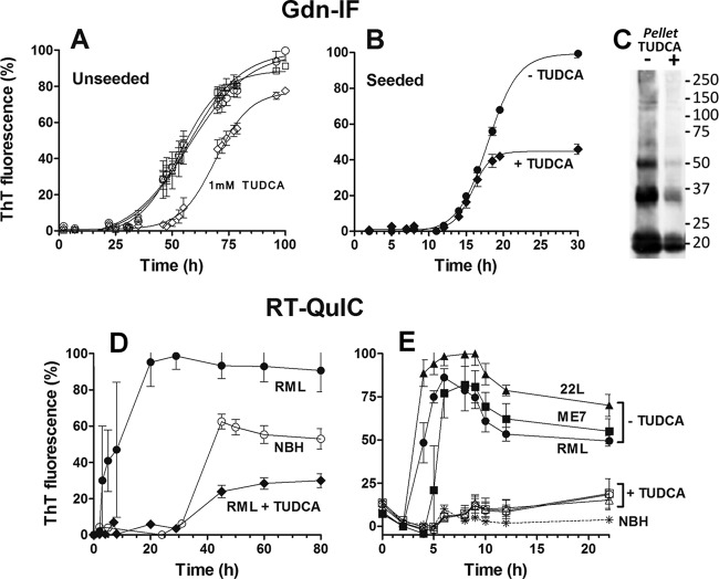
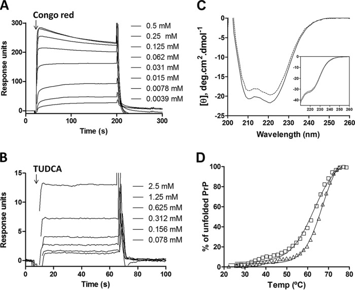
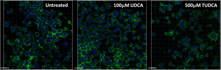
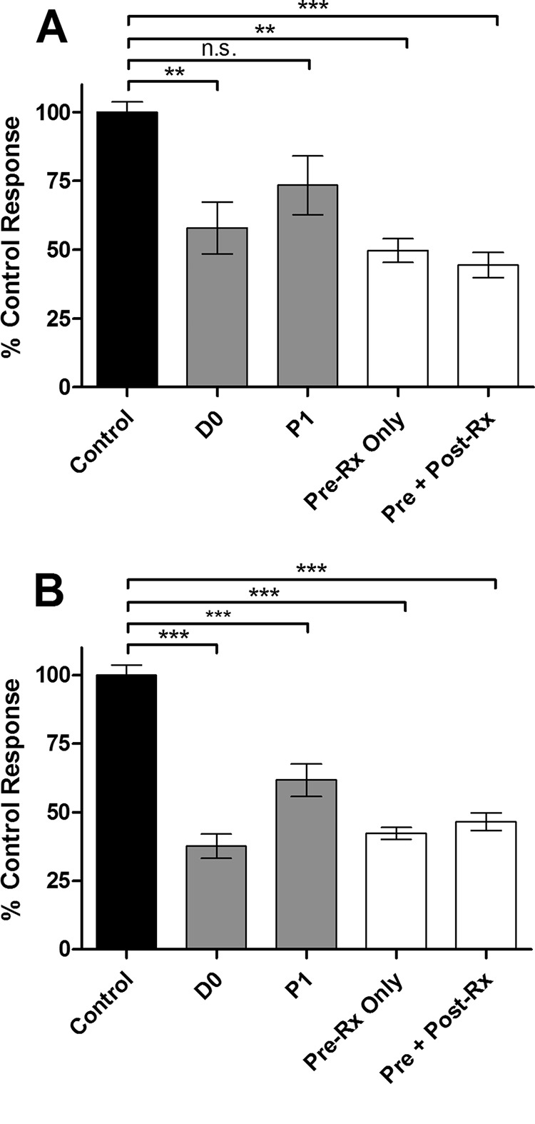
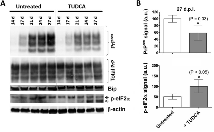
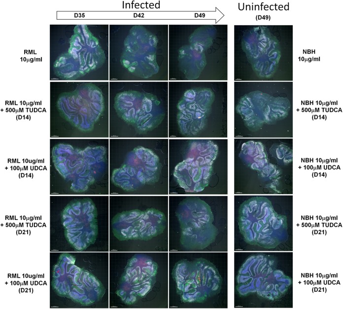
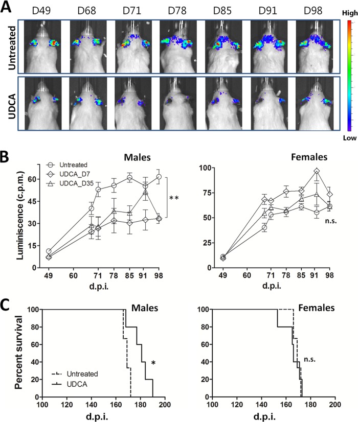
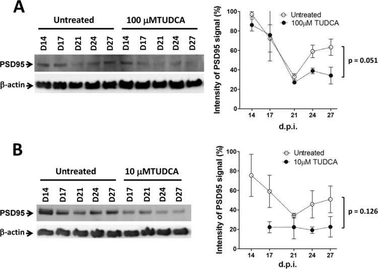
Prion diseases are fatal neurodegenerative disorders associated with the conversion of cellular prion protein (PrPC) into its aberrant infectious form (PrPSc). There is no treatment available for these diseases. The bile acids tauroursodeoxycholic acid(TUDCA) and ursodeoxycholic acid (UDCA) have been recently shown to be neuroprotective in other protein misfolding disease models, including Parkinson’s, Huntington’s and Alzheimer’s diseases, and also in humans with amyotrophic lateral sclerosis.Here, we studied the therapeutic efficacy of these compounds in prion disease. We demonstrated that TUDCA and UDCA substantially reduced PrP conversion in cell-free aggregation assays, as well as in chronically and acutely infected cell cultures. This effect was mediated through reduction of PrPSc seeding ability, rather than an effect on PrPC. We also demonstrated the ability of TUDCA and UDCA to reduce neuronal loss in prion-infected cerebellar slice cultures. UDCA treatment reduced astrocytosis and prolonged survival in RML prion-infected mice. Interestingly, these effects were limited to the males, implying a gender-specific difference in drug metabolism. Beyond effects on PrPSc, we found that levels of phosphorylated eIF2 were increased at early time points, with correlated reductions in postsynaptic density protein 95. As demonstrated for other neurodegenerative diseases, we now show that TUDCA and UDCA may have a therapeutic role in prion diseases, with effects on both prion conversion and neuroprotection. Our findings, together with the fact that these natural compounds are orally bioavailable, permeable to the blood-brain barrier, and U.S. Food and Drug Administration-approved for use in humans, make these compounds promising alternatives for the treatment of prion diseases.

Importance: Prion diseases are fatal neurodegenerative diseases that are transmissible to humans and other mammals. There are no disease-modifying therapies available, despite decades of research. Treatment targets have included inhibition of protein accumulation,clearance of toxic aggregates, and prevention of downstream neurodegeneration. No one target may be sufficient; rather, compounds which have a multimodal mechanism, acting on different targets, would be ideal. TUDCA and UDCA are bile acids that may fulfill this dual role. Previous studies have demonstrated their neuroprotective effects in several neurodegenerative disease models, and we now demonstrate that this effect occurs in prion disease, with an added mechanistic target of upstream prion seeding. Importantly, these are natural compounds which are orally bioavailable, permeable to the blood-brain barrier, and U.S.Food and Drug Administration-approved for use in humans with primary biliary cirrhosis. They have recently been proven efficacious in human amyotrophic lateral sclerosis. Therefore, these compounds are promising options for the treatment of prion diseases.


Figures

FIG 1
FIG 1
Effect of TUDCA on PrP(90-231) fibril formation, as monitored by ThT fluorescence. (A and B) PrP(90-231) at 22 μM was incubated under Gdn-IF conditions (50 mM phosphate buffer [pH 7.0] and 2 M GdnHCl at 37°C and 500 rpm) in the absence (A) or presence (B) of seeds. (A) Unseeded PrP(90-231) was aggregated alone (○) or with the addition of 0.01 mM (□), 0.1 mM (△), and 1 mM (♢) TUDCA. B, PrP(90-231) seeded with 0.00025% (wt/vol) preformed fibrils was aggregated with (◆) or without (●) the addition of 1 mM TUDCA. (C) Immunoblot of total aggregated PrP(90-231) formed from under seeded conditions (corresponding to the kinetics curves from panel B) with (+) or without (−) 1 mM TUDCA. The total volume of the aggregation reaction was centrifuged at 10,000 × g for 30 min, and the pellet was resuspended in sample buffer and analyzed by immunoblotting with SAF83. (D and E) PrP(90-231) at 10 μM was incubated under RT-QuIC conditions (10 mM phosphate buffer [pH 7.4], 130 mM NaCl, 10 μM EDTA, and 0.002% SDS at 41°C and 900 rpm) in the presence of normal mouse brain homogenate (NBH) or prion-infected brain homogenate. (D) PrP(90-231) was aggregated in the presence of 0.0007% (wt/vol) NBH (○) or RML brain homogenate with (◆) or without (●) 1 mM TUDCA. (E) PrP(90-231) was unseeded (asterisk) or seeded with 0.0007% (wt/vol) RML (circles), ME7 (squares) and 22L (triangle) strains with (empty symbols) or without (filled symbols) 1 mM TUDCA. Each kinetic was performed in triplicate, and the means ± the standard errors of the mean (SEM) are shown.
FIG 2
FIG 2
Interaction of TUDCA with PrP(90-231) as measured by SPR and CD analyses. (A and B) Sensograms of His-tagged PrP(90-231), captured on an NTA-sensor chip, interacting with serial dilutions of Congo red (A) or TUDCA (B). (C) Ellipticity of monomeric PrP(90-231) in the absence (dashed line) or presence (filled line) of 1 mM TUDCA determined under the RT-QuIC and Gdn-IF (inset) buffer conditions. Ten spectra were recorded at 25°C and averaged. (D) Thermal denaturation of PrP(90-231) in the absence (triangles) or presence (squares) of 1 mM TUDCA was monitored by measuring ellipticity at 222 nm.
FIG 3
FIG 3
TUDCA and UDCA treatment of N2a cells chronically infected with RML (ScN2a cells). (A) Representative immunoblots of PrPres in ScN2a cells through six passages (3 to 4 days each) in the presence of 100 μM TUDCA (top panel) or 100 μM UDCA (bottom panel). Cells were harvested and treated with 20 μg of PK/ml for 1 h at 37°C. (B) Mean ± the SE densitometries of PrPres from two experiments (*, P < 0.05 [statistically significant difference from control]). TUDCA, diamonds; UDCA, triangles. Signal was normalized to untreated control cells (P0).
FIG 4
FIG 4
Effect of bile acids on PrPC distribution and cell morphology. N2a cells were exposed to 100 μM UDCA or 500 μM TUDCA for 24 h and then fixed, permeabilized, and labeled with SAF83 for PrPC (green) and DAPI (blue). Images were obtained at ×20 using a Zeiss LSM. Scale bars, 40 μm.
FIG 5
FIG 5
TUDCA and UDCA treatment of L929 cells acutely infected with RML (scrapie cell assay). Levels of PrPres after three passages (5 days each) in a scrapie cell assay in the presence of 100 μM TUDCA (A) or UDCA (B). The cells were treated immediately after RML infection (D0), starting after the first passage (P1), or were exposed to infected brain homogenate preincubated with bile acid (Pre-Rx), followed by no further addition (Pre-Rx only) or the addition of 100 μM TUDCA at passage 1 (Pre + Post-Rx). The readout was normalized to untreated infected control wells. The data represent mean ± the SEM. n = 8 for D0 and P1; n = 4 for Pre-Rx only and Pre + Post-Rx. ***, P < 0.001; **, P < 0.01 (statistically significant difference from the control). n.s., P > 0.05.
FIG 6
FIG 6
TUDCA treatment of RML-infected cerebellar slice cultures. (A) Immunoblot for PrPres, total PrP, BiP, and p-eIF2α of RML-infected cerebellar slice cultures left untreated or treated with 100 μM TUDCA starting immediately after infection. Samples were harvested at days 14, 17, 24, and 27 postinfection. (B) Quantification of PrPres and p-eIF2α signals from three independent samples harvested at 27 dpi. The data represent means ± the SEM. *, P < 0.05 (statistically significant difference from the control).
FIG 7
FIG 7
Bile acid-induced neuroprotection ex vivo. Cerebellar slices were stained with antibodies to Calbindin (green) and NeuN (white), in addition to TUNEL (red) and DAPI (blue) counterstains. n = 3 (representative images are shown). RML-infected slices were treated with 500 μM TUDCA or 100 μM starting at day 14 (D14) or 21 (D21) postinfection. Slices were collected at days 35 (D35), 42 (D42), and 49 (D49), stained, and analyzed by confocal microscopy. Prion infection elicited a significant loss of granule cell neurons (NeuN+) and Purkinje cells (Calbindin+). Uninfected controls were inoculated with normal brain homogenate (NBH) and were either left untreated or treated with TUDCA or UDCA starting at day 14 or 21.
FIG 8
FIG 8
Bioluminescence and survival curves of RML-infected Tg(Gfap-luc) and C57BL/6 mice treated with UDCA. Tg(Gfap-luc) mice were intracerebrally inoculated with 1% RML-infected brain homogenate and treated with 0.03% (wt/vol) UDCA, whereas C57BL/6 mice were intracerebrally inoculated with 0.1% RML-infected brain homogenate and treated with 0.05% (wt/vol) UDCA. (A) Representative scans of one infected untreated male mouse (top panels) and one infected treated male mouse (bottom panel). Treatment started at 7 dpi. Scans were taken at 49, 68, 71, 78, 85, 91, and 98 dpi. (B) Bioluminescence quantification from the brains of male (left) and female (right) infected Tg(Gfap-luc) mice untreated (circles) and treated starting at 7 dpi (diamonds) or 35 dpi (triangles). The data show means ± the SEM (n = 6 for control and n = 4 for mice treated at day 35 and females treated at day 7; n = 3 for males treated at day 7; **, P < 0.01). (C) Kaplan-Meier plots for survival time of male (left) and female (right) infected C57BL/6 mice untreated (dashed curve) and treated starting at day 35 (solid curve). Log-rank (Mantel-Cox) and Gehan-Breslow-Wilcoxon tests were applied for statistical analysis obtaining P values of 0.021 and 0.039, respectively (n = 6 for control [3 males, 3 females]; n = 5 for treated males; n = 5 for treated females; *, P < 0.05).
FIG 9
FIG 9
Effect of TUDCA on PSD95 levels of infected cerebellar slices. (A and B) Western blots and densitometric analysis of PSD95 expression levels in POSCA untreated and treated with 100 μM TUDCA starting at day 0 (A) or 10 μM TUDCA starting at day 7 (B) postinfection. Samples were harvested at days 14, 17, 21, 24, and 27. The data represent means ± the SEM (n = 3 for untreated and n = 2 for treated samples).

References

    1. Sim VL. 2012. Prion disease: chemotherapeutic strategies. Infect Disord Drug Targets 12:144–160. doi:10.2174/187152612800100161. - DOI - PubMed
    1. Cortez L, Sim V. 2014. The therapeutic potential of chemical chaperones in protein folding diseases. Prion 8:197–202. doi:10.4161/pri.28938. - DOI - PMC - PubMed
    1. Geier A, Wagner M, Dietrich CG, Trauner M. 2007. Principles of hepatic organic anion transporter regulation during cholestasis, inflammation, and liver regeneration. Biochim Biophys Acta 1773:283–308. doi:10.1016/j.bbamcr.2006.04.014. - DOI - PubMed
    1. Rodrigues CM, Fan G, Wong PY, Kren BT, Steer CJ. 1998. Ursodeoxycholic acid may inhibit deoxycholic acid-induced apoptosis by modulating mitochondrial transmembrane potential and reactive oxygen species production. Mol Med 4:165–178. doi:10.1007/s0089480040165. - DOI - PMC - PubMed
    1. Rodrigues CM, Fan G, Ma X, Kren BT, Steer CJ. 1998. A novel role for ursodeoxycholic acid in inhibiting apoptosis by modulating mitochondrial membrane perturbation. J Clin Invest 101:2790–2799. doi:10.1172/JCI1325. - DOI - PMC - PubMed

Publication types

LinkOut - more resources