

Transcriptome Profiling of the Virus-Induced Innate Immune Response in Pteropus vampyrus and Its Attenuation by Nipah Virus Interferon Antagonist Functions
- PMID: 25972557
- PMCID: PMC4505658
- DOI: 10.1128/JVI.00302-15
Transcriptome Profiling of the Virus-Induced Innate Immune Response in Pteropus vampyrus and Its Attenuation by Nipah Virus Interferon Antagonist Functions
Abstract
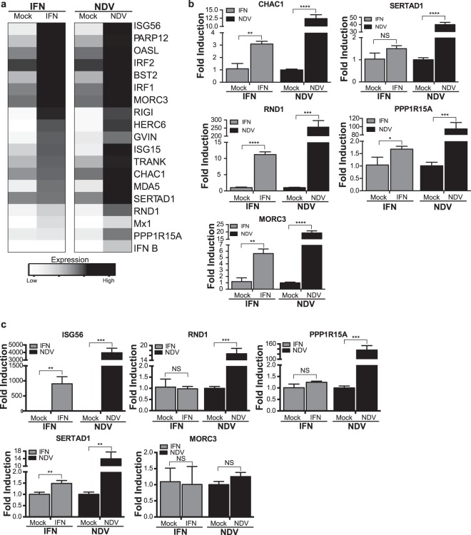
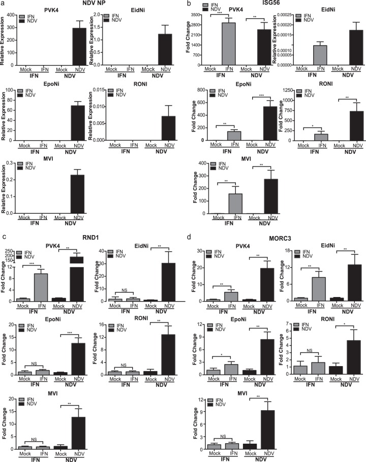
Bats are important reservoirs for several viruses, many of which cause lethal infections in humans but have reduced pathogenicity in bats. As the innate immune response is critical for controlling viruses, the nature of this response in bats and how it may differ from that in other mammals are of great interest. Using next-generation transcriptome sequencing (mRNA-seq), we profiled the transcriptional response of Pteropus vampyrus bat kidney (PVK) cells to Newcastle disease virus (NDV), an avian paramyxovirus known to elicit a strong innate immune response in mammalian cells. The Pteropus genus is a known reservoir of Nipah virus (NiV) and Hendra virus (HeV). Analysis of the 200 to 300 regulated genes showed that genes for interferon (IFN) and antiviral pathways are highly upregulated in NDV-infected PVK cells, including genes for beta IFN, RIG-I, MDA5, ISG15, and IRF1. NDV-infected cells also upregulated several genes not previously characterized to be antiviral, such as RND1, SERTAD1, CHAC1, and MORC3. In fact, we show that MORC3 is induced by both IFN and NDV infection in PVK cells but is not induced by either stimulus in human A549 cells. In contrast to NDV infection, HeV and NiV infection of PVK cells failed to induce these innate immune response genes. Likewise, an attenuated response was observed in PVK cells infected with recombinant NDVs expressing the NiV IFN antagonist proteins V and W. This study provides the first global profile of a robust virus-induced innate immune response in bats and indicates that henipavirus IFN antagonist mechanisms are likely active in bat cells.
Importance: Bats are the reservoir host for many highly pathogenic human viruses, including henipaviruses, lyssaviruses, severe acute respiratory syndrome coronavirus, and filoviruses, and many other viruses have also been isolated from bats. Viral infections are reportedly asymptomatic or heavily attenuated in bat populations. Despite their ecological importance to viral maintenance, research into their immune system and mechanisms for viral control has only recently begun. Nipah virus and Hendra virus are two paramyxoviruses associated with high mortality rates in humans and whose reservoir is the Pteropus genus of bats. Greater knowledge of the innate immune response of P. vampyrus bats to viral infection may elucidate how bats serve as a reservoir for so many viruses.
Copyright © 2015, American Society for Microbiology. All Rights Reserved.
Figures
Similar articles
-
Nipah and Hendra Virus Nucleoproteins Inhibit Nuclear Accumulation of Signal Transducer and Activator of Transcription 1 (STAT1) and STAT2 by Interfering with Their Complex Formation.J Virol. 2017 Oct 13;91(21):e01136-17. doi: 10.1128/JVI.01136-17. Print 2017 Nov 1. J Virol. 2017. PMID: 28835499 Free PMC article.
-
Understanding the interaction between henipaviruses and their natural host, fruit bats: Paving the way toward control of highly lethal infection in humans.Int Rev Immunol. 2017 Mar 4;36(2):108-121. doi: 10.1080/08830185.2016.1255883. Epub 2017 Jan 6. Int Rev Immunol. 2017. PMID: 28060559
-
Pteropid bats are confirmed as the reservoir hosts of henipaviruses: a comprehensive experimental study of virus transmission.Am J Trop Med Hyg. 2011 Nov;85(5):946-51. doi: 10.4269/ajtmh.2011.10-0567. Am J Trop Med Hyg. 2011. PMID: 22049055 Free PMC article.
-
Henipaviruses: an updated review focusing on the pteropid reservoir and features of transmission.Zoonoses Public Health. 2013 Feb;60(1):69-83. doi: 10.1111/j.1863-2378.2012.01501.x. Epub 2012 Jun 18. Zoonoses Public Health. 2013. PMID: 22709528 Review.
-
Nipah and hendra virus interactions with the innate immune system.Curr Top Microbiol Immunol. 2012;359:123-52. doi: 10.1007/82_2012_209. Curr Top Microbiol Immunol. 2012. PMID: 22491899 Review.
Cited by
-
Genomic profiling of Nipah virus using NGS driven RNA-Seq expression data.Bioinformation. 2019 Dec 31;15(12):853-862. doi: 10.6026/97320630015853. eCollection 2019. Bioinformation. 2019. PMID: 32256005 Free PMC article.
-
An intersectional analysis of LncRNAs and mRNAs reveals the potential therapeutic targets of Bi Zhong Xiao Decoction in collagen-induced arthritis rats.Chin Med. 2022 Sep 16;17(1):110. doi: 10.1186/s13020-022-00670-z. Chin Med. 2022. PMID: 36109779 Free PMC article.
-
Bats and Coronaviruses.Viruses. 2019 Jan 9;11(1):41. doi: 10.3390/v11010041. Viruses. 2019. PMID: 30634396 Free PMC article. Review.
-
Coronavirus, the King Who Wanted More Than a Crown: From Common to the Highly Pathogenic SARS-CoV-2, Is the Key in the Accessory Genes?Front Microbiol. 2021 Jul 14;12:682603. doi: 10.3389/fmicb.2021.682603. eCollection 2021. Front Microbiol. 2021. PMID: 34335504 Free PMC article. Review.
-
Natural genetic variation in Drosophila melanogaster reveals genes associated with Coxiella burnetii infection.Genetics. 2021 Mar 31;217(3):iyab005. doi: 10.1093/genetics/iyab005. Genetics. 2021. PMID: 33789347 Free PMC article.
References
-
- Halpin K, Hyatt AD, Fogarty R, Middleton D, Bingham J, Epstein JH, Rahman SA, Hughes T, Smith C, Field HE, Daszak P. 2011. Pteropid bats are confirmed as the reservoir hosts of henipaviruses: a comprehensive experimental study of virus transmission. Am J Trop Med Hyg 85:946–951. doi:10.4269/ajtmh.2011.10-0567. - DOI - PMC - PubMed
-
- Halpin K, Young PL, Field HE, Mackenzie JS. 2000. Isolation of Hendra virus from pteropid bats: a natural reservoir of Hendra virus. J Gen Virol 81:1927–1932. - PubMed
Publication types
MeSH terms
Substances
Grants and funding
LinkOut - more resources
Full Text Sources
Other Literature Sources
Research Materials
Miscellaneous
