Review
. 2015 Apr 28:9:60.
doi: 10.3389/fnsys.2015.00060. eCollection 2015.

Visual system plasticity in mammals: the story of monocular enucleation-induced vision loss

Affiliations
Review

Visual system plasticity in mammals: the story of monocular enucleation-induced vision loss

Julie Nys et al. Front Syst Neurosci. .

Abstract

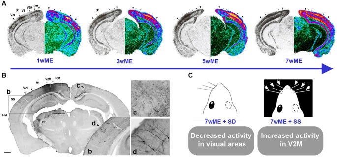
The groundbreaking work of Hubel and Wiesel in the 1960's on ocular dominance plasticity instigated many studies of the visual system of mammals, enriching our understanding of how the development of its structure and function depends on high quality visual input through both eyes. These studies have mainly employed lid suturing, dark rearing and eye patching applied to different species to reduce or impair visual input, and have created extensive knowledge on binocular vision. However, not all aspects and types of plasticity in the visual cortex have been covered in full detail. In that regard, a more drastic deprivation method like enucleation, leading to complete vision loss appears useful as it has more widespread effects on the afferent visual pathway and even on non-visual brain regions. One-eyed vision due to monocular enucleation (ME) profoundly affects the contralateral retinorecipient subcortical and cortical structures thereby creating a powerful means to investigate cortical plasticity phenomena in which binocular competition has no vote.In this review, we will present current knowledge about the specific application of ME as an experimental tool to study visual and cross-modal brain plasticity and compare early postnatal stages up into adulthood. The structural and physiological consequences of this type of extensive sensory loss as documented and studied in several animal species and human patients will be discussed. We will summarize how ME studies have been instrumental to our current understanding of the differentiation of sensory systems and how the structure and function of cortical circuits in mammals are shaped in response to such an extensive alteration in experience. In conclusion, we will highlight future perspectives and the clinical relevance of adding ME to the list of more longstanding deprivation models in visual system research.

Keywords: age; cortical plasticity; deafferentation; deprivation; multimodal; reorganization; visual system.


Figures

Figure 1
Figure 1
Spatiotemporal reactivation of the contralateral visual cortex by visual and cross-modal inputs after monocular enucleation (ME) in adult mice. (A) Layer- and time-specific recovery of neuronal activity in the left visual cortex subsequent to removal of the right eye at an adult age of P120 is illustrated. Molecular activity profiles of the visual cortex have been assessed by the zif268 mRNA expression analysis around Bregma level −3.40 mm. For each section, the original autoradiogram displaying the deprived (left) visual cortex is shown in gray and its matching pseudo-colored mirror image. The medial and lateral extent of the left visual cortex is marked by the two large arrowheads whereas small arrowheads delineate the interareal boundaries. The activity in the central binocular cortex starts to expand supragranularly (asterisk first and second panel) between 1 and 3 weeks post-ME. Between 3 and 5 weeks, infragranular layers also start to show increased reactivation. (B) Anterograde and retrograde transport of fluororuby upon injection in V2M of a 7wME mouse. b: Detail of Fluororuby signal at the location of somatosensory cortex: tracer is transported in an anterograde way to axon terminals in layers V and VI, while supragranular layers II/III contain retrogradely labeled cell bodies and dendrites. c: Detail of anterogradely labeled fibers in contralateral V2M. d: Detail of retrogradely labeled pyramidal cells in layers II and III of ipsilateral/adjacent somatosensory cortex. (C) Subsequent whisker manipulations in 7wME mice were employed in order to verify the functional relevance of the intermodal connections in the ME-induced reactivation profile. Somatosensory deprivation (SD) by trimming the right-side vibrissae results in decreased visual cortex activity whereas somatosensory stimulation (SS) through exposure to toys and novel objects in the dark increased activity, especially in V2M. Adapted from Van Brussel et al. (2011).

References

    1. Aerts J., Nys J., Arckens L. (2014a). A highly reproducible and straightforward method to perform in vivo ocular enucleation in the mouse after eye opening. J. Vis. Exp. 6:e51936. 10.3791/51936 - DOI - PMC - PubMed
    1. Aerts J., Nys J., Moons L., Hu T. T., Arckens L. (2014b). Altered neuronal architecture and plasticity in the visual cortex of adult MMP-3 deficient mice. Brain Struct. Funct. [Epub ahead of print]. 10.1007/s00429-014-0819-4 - DOI - PubMed
    1. Allman B. L., Keniston L. P., Meredith M. A. (2009a). Adult deafness induces somatosensory conversion of ferret auditory cortex. Proc. Natl. Acad. Sci. U S A 106, 5925–5930. 10.1073/pnas.0809483106 - DOI - PMC - PubMed
    1. Allman B. L., Keniston L. P., Meredith M. A. (2009b). Not just for bimodal neurons anymore: the contribution of unimodal neurons to cortical multisensory processing. Brain Topogr. 21, 157–167. 10.1007/s10548-009-0088-3 - DOI - PMC - PubMed
    1. Andermann M. L., Kerlin A. M., Roumis D. K., Glickfeld L. L., Reid R. C. (2011). Functional specialization of mouse higher visual cortical areas. Neuron 72, 1025–1039. 10.1016/j.neuron.2011.11.013 - DOI - PMC - PubMed

LinkOut - more resources