. 2015 Apr 28:6:116.
doi: 10.3389/fphys.2015.00116. eCollection 2015.

Pseudo-acetylation of K326 and K328 of actin disrupts Drosophila melanogaster indirect flight muscle structure and performance

Affiliations

Pseudo-acetylation of K326 and K328 of actin disrupts Drosophila melanogaster indirect flight muscle structure and performance

Meera C Viswanathan et al. Front Physiol. .

Abstract

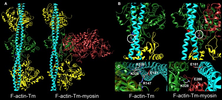
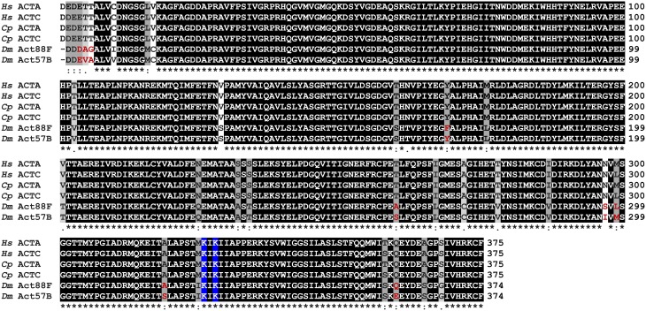
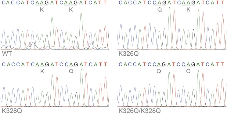
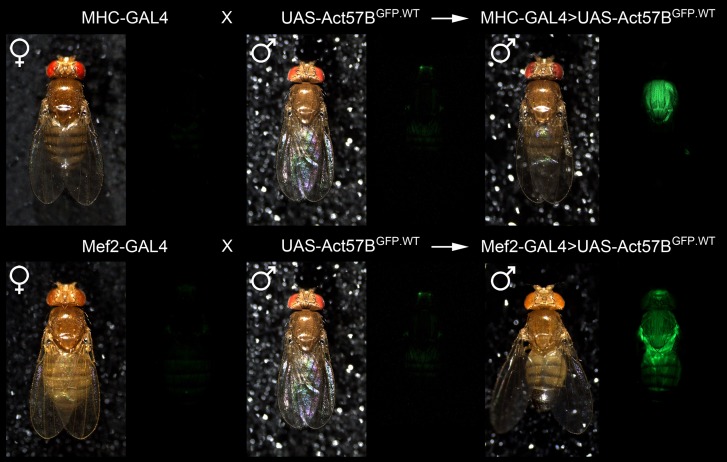
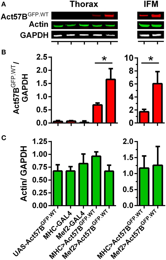
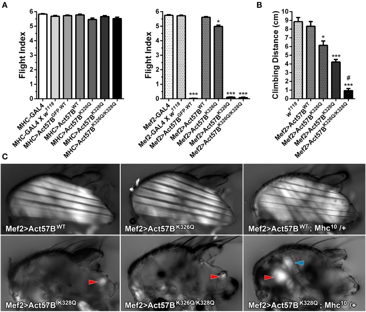
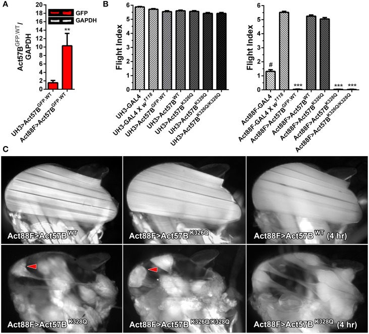
In striated muscle tropomyosin (Tm) extends along the length of F-actin-containing thin filaments. Its location governs access of myosin binding sites on actin and, hence, force production. Intermolecular electrostatic associations are believed to mediate critical interactions between the proteins. For example, actin residues K326, K328, and R147 were predicted to establish contacts with E181 of Tm. Moreover, K328 also potentially forms direct interactions with E286 of myosin when the motor is strongly bound. Recently, LC-MS/MS analysis of the cardiac acetyl-lysine proteome revealed K326 and K328 of actin were acetylated, a post-translational modification (PTM) that masks the residues' inherent positive charges. Here, we tested the hypothesis that by removing the vital actin charges at residues 326 and 328, the PTM would perturb Tm positioning and/or strong myosin binding as manifested by altered skeletal muscle function and structure in the Drosophila melanogaster model system. Transgenic flies were created that permit tissue-specific expression of K326Q, K328Q, or K326Q/K328Q acetyl-mimetic actin and of wild-type actin via the UAS-GAL4 bipartite expression system. Compared to wild-type actin, muscle-restricted expression of mutant actin had a dose-dependent effect on flight ability. Moreover, excessive K328Q and K326Q/K328Q actin overexpression induced indirect flight muscle degeneration, a phenotype consistent with hypercontraction observed in other Drosophila myofibrillar mutants. Based on F-actin-Tm and F-actin-Tm-myosin models and on our physiological data, we conclude that acetylating K326 and K328 of actin alters electrostatic associations with Tm and/or myosin and thereby augments contractile properties. Our findings highlight the utility of Drosophila as a model that permits efficient targeted design and assessment of molecular and tissue-specific responses to muscle protein modifications, in vivo.

Keywords: acetylation; muscle contraction; myosin; post-translational modification; tropomyosin.


Figures

Figure 1
Figure 1
Critical electrostatic F-actin-Tm and F-actin-Tm-myosin interactions. (A) Molecular models showing the location of tropomyosin (Tm) (blue) on actin (yellow and green) in the absence and presence of the myosin head (S1) (red) bound in rigor. The F-actin-Tm and rigor F-actin-Tm-myosin structures are based on those generated by Li et al. (2011) and Behrmann et al. (2012) respectively. (B) Enlarged views illustrate critical electrostatic associations between actin and Tm in the absence or presence of S1. K328 on actin (circled) interacts with E181 of Tm in the absence of myosin (left) and with E286 of myosin when S1 is bound in rigor (right). Note the azimuthal movement of Tm across F-actin. (C) Projected and enlarged views highlight vital electrostatic interactions of actin residues R147, K326, and K328 with E181 of Tm, in the absence of myosin, and of K328 of actin and E286 of S1 when myosin is bound in rigor. These associations are likely critical for thin filament and muscle function.
Figure 2
Figure 2
Multiple sequence alignment of actin isoforms. Multiple sequence alignment of skeletal and cardiac actin from Homo sapiens (Hs), Cavia porcellus (Cp), and Drosophila melanogaster (Dm) reveals highly conserved proteins. ACTA, skeletal muscle actin; ACTC, cardiac muscle actin; Act88F, Drosophila indirect flight muscle actin; Act57B, Drosophila cardiac actin. Residues are shaded based on degree of conservation. An (*) indicates positions that have identical residues, a (:) indicates substitution with high structural similarity, and a (.) indicates substitution low structural similarity.
Figure 3
Figure 3
Confirmation of transgenic actin transcription. Sequence chromatograms of an amplified stretch of Act57B cDNA revealed transcription of the UAS-Act57B transgenes in the thoracic musculature of Mef2-GAL4> UAS-Act57B transgenic flies. The chromatograms also confirmed the presence and expression of K326Q, K328Q, or K326Q/K328Q actin mutations (identified by the AAG → CAG nucleotide transversion) in the sequenced Act57B cDNA fragments.
Figure 4
Figure 4
Confirmation of muscle-restricted gene expression. Virgin female flies expressing either the muscle-specific MHC-GAL4 or the Mef2-GAL4 driver were mated with male flies carrying the UAS-Act57BGFP.WT construct. Background fluorescence coming from the musculature of the parental lines was minimal. However, fluorescence emitted from the musculature of progeny, which inherit both a GAL4 driver and the UAS-construct, was readily observed in both genotypes, confirming tissue-specific expression of transgenic Act57B actin.
Figure 5
Figure 5
Mef2-GAL4 drives higher expression levels of transgenic actin relative to MHC-GAL4. Quantitative western blot analysis of steady-state Act57BGFP.WT and total actin was performed on thoraces and IFMs of Mef2-GAL4> UAS-Act57BGFP.WT and MHC-GAL4> UAS-Act57BGFP.WT flies and of control flies two days after eclosion. (A) Representative western blots showing elevated thoracic (left) and IFM (right) levels of Act57BGFP.WT (probed with an anti-GFP primary antibody) when driven by Mef2-GAL4 compared to MHC-GAL4. Actin and GAPDH (probed with anti-actin and anti-GAPDH antibodies) abundance appeared relatively consistent among genotypes. The GFP (B) and actin (C) intensities were measured, normalized to that of GAPDH for five thoracic samples with six technical replicates each and for eight IFM samples with four technical replicates each, and averaged for each genotype. Mef2-GAL4 drove significantly higher amounts of transgenic actin relative to MHC-GAL4 (*P < 0.05).
Figure 6
Figure 6
Excessive expression levels of GFP-tagged or acetyl-mimetic actin disrupt muscle function and structure. (A) Flight indices of control and of MHC-GAL4> or Mef2-GAL4> UAS-Act57BGFP.WT, UAS-Act57BWT, UAS-Act57BK326Q, UAS-Act57BK328Q, or UAS-Act57BK326Q/K328Q transgenic Drosophila. MHC-GAL4 “low-dose”-driven UAS-Act57B constructs did not affect flight ability in any transgenic line. “High-dose” expression of UAS-Act57BGFP.WT by Mef2-GAL4 abolished flight whereas UAS-Act57BWT transgene expression had no effect on flight performance. Mef2-GAL4-driven expression of UAS-Act57BK326Q caused a slight but significant reduction in flight ability (*P < 0.05 compared to controls), while expression of UAS-Act57BK328Q or UAS-Act57BK326Q/K328Q completely eliminated flight (***P < 0.001 compared to controls). (B) Effects of pseudo-acetylation on climbing ability. Pseudo-acetylated K326Q actin showed the least and K326Q/K328Q actin the most damaging effects, which illustrates the PTM can also influence performance of non-fibrillar muscle (*P < 0.05, ***P < 0.001 compared to controls; #P < 0.01 compared to Mef2-GAL4> UAS-Act57BK328Q) (C) Polarized light micrographs of IFM from Mef2-GAL4> UAS-Act57BWT, UAS-Act57BK326Q, UAS-Act57BK328Q, or UAS-Act57BK326Q/K328Q flies. Mef2-GAL4> UAS-Act57BK326Q IFM appeared indistinguishable from Mef2-GAL4> UAS-Act57BWT control. Mef2-GAL4-mediated expression of UAS-Act57BK328Q and UAS-Act57BK326Q/K328Q, however, resulted in a phenotype consistent with severe hypercontraction. Minor traces of birefringent material, assumed to be IFM remnants (red arrowheads), were occasionally observed. A single copy of Mhc10, the IFM-specific myosin null allele, had no influence on gross muscle morphology in Mef2-GAL4> UAS-Act57BWT; Mhc10/+ thoraces. Mef2-GAL4> UAS-Act57BK328Q; Mhc10/+ Drosophila displayed increased abundance of birefringent thoracic musculature (red arrowhead) relative to Mef2-GAL4> UAS-Act57BK328Q flies. The blue arrowhead indicates the tergal depressor of trochanter (jump) muscle. These findings are consistent with previous studies that demonstrated reduced MHC partially suppresses fiber destruction and they suggest that IFM expressing Mef2-GAL4-driven UAS-Act57BK328Q requires relatively little myosin to hypercontract.
Figure 7
Figure 7
Disproportionately high expression of UAS-Act57BK328Q via the Act88F-GAL4 IFM-specific driver induces hypercontraction. (A) Quantitative western blot analysis of Act57BGFP.WT abundance driven by UH3-GAL4 vs. Act88F-GAL4. GFP intensities were normalized to that of GAPDH and averaged for eight IFM samples with four technical replicates each. Act88F-GAL4 drove significantly higher amounts of transgenic actin relative to UH3-GAL4 (**P < 0.01). (B) Flight indices of UH3-GAL4> and Act88F-GAL4> UAS-Act57BGFP.WT, UAS-Act57BWT, UAS-Act57BK326Q, UAS-Act57BK328Q, and UAS-Act57BK326Q/K328Q transgenic Drosophila. UH3-GAL4, and “low dose” expression of all UAS-Act57B actin constructs by the driver, had no effect on flight. Act88F-GAL4 Drosophila exhibited significantly reduced flight performance relative to female progeny of Act88F-GAL4 x w1118 (#P < 0.001). The latter demonstrated wild-type-like flight ability. “High-dose” expression of UAS-Act57BGFP.WT by Act88F-GAL4 eliminated flight whereas UAS-Act57BWT transgene expression had no effect on flight performance. “High dose” expression of UAS-Act57BK326Q reduced flight ability, an effect which approached statistical significance. Act88F-GAL4> UAS-Act57BK328Q and UAS-Act57BK326Q/K328Q Drosophila were flightless (***P < 0.001). (C) Fluorescent images of dorsal longitudinal IFMs from two-day-old Act88F-GAL4> UAS-Act57BWT, UAS-Act57BK326Q, UAS-Act57BK328Q, and UAS-Act57BK326Q/K328Q flies. Act88F-GAL4> UAS-Act57BK326Q IFMs were indistinguishable from Act88F-GAL4> UAS-Act57BWT control IFMs. Act88F-GAL4> UAS-Act57BK328Q and UAS-Act57BK326Q/K328Q Drosophila, however, displayed hypercontracted IFM with separated and bunched fibers (red arrowheads) at attachment sites. IFMs from young (four hour old) UAS-Act57BK328Q-expressing flies had a considerably less severe phenotype with minimal thinning and separation of the fibers.

Similar articles

Cited by

References

    1. Agnetti G., Husberg C., van Eyk J. E. (2011). Divide and conquer the application of organelle proteomics to heart failure. Circ. Res. 108, 512–526. 10.1161/CIRCRESAHA.110.226910 - DOI - PMC - PubMed
    1. An H. S., Mogami K. (1996). Isolation of 88F actin mutants of Drosophila melanogaster and possible alterations in the mutant actin structures. J. Mol. Biol. 260, 492–505. 10.1006/jmbi.1996.0417 - DOI - PubMed
    1. Barua B., Fagnant P. M., Winkelmann D. A., Trybus K. M., Hitchcock-Degregori S. E. (2013). A periodic pattern of evolutionarily conserved basic and acidic residues constitutes the binding interface of actin-tropomyosin. J. Biol. Chem. 288, 9602–9609. 10.1074/jbc.M113.451161 - DOI - PMC - PubMed
    1. Barua B., Pamula M. C., Hitchcock-Degregori S. E. (2011). Evolutionarily conserved surface residues constitute actin binding sites of tropomyosin. Proc. Natl. Acad. Sci. U.S.A. 108, 10150–10155. 10.1073/pnas.1101221108 - DOI - PMC - PubMed
    1. Barua B., Winkelmann D. A., White H. D., Hitchcock-Degregori S. E. (2012). Regulation of actin-myosin interaction by conserved periodic sites of tropomyosin. Proc. Natl. Acad. Sci. U.S.A. 109, 18425–18430. 10.1073/pnas.1212754109 - DOI - PMC - PubMed

LinkOut - more resources