

Audiovisual integration of speech in a patient with Broca's Aphasia
- PMID: 25972819
- PMCID: PMC4411977
- DOI: 10.3389/fpsyg.2015.00435
Audiovisual integration of speech in a patient with Broca's Aphasia
Abstract
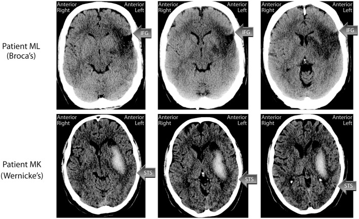
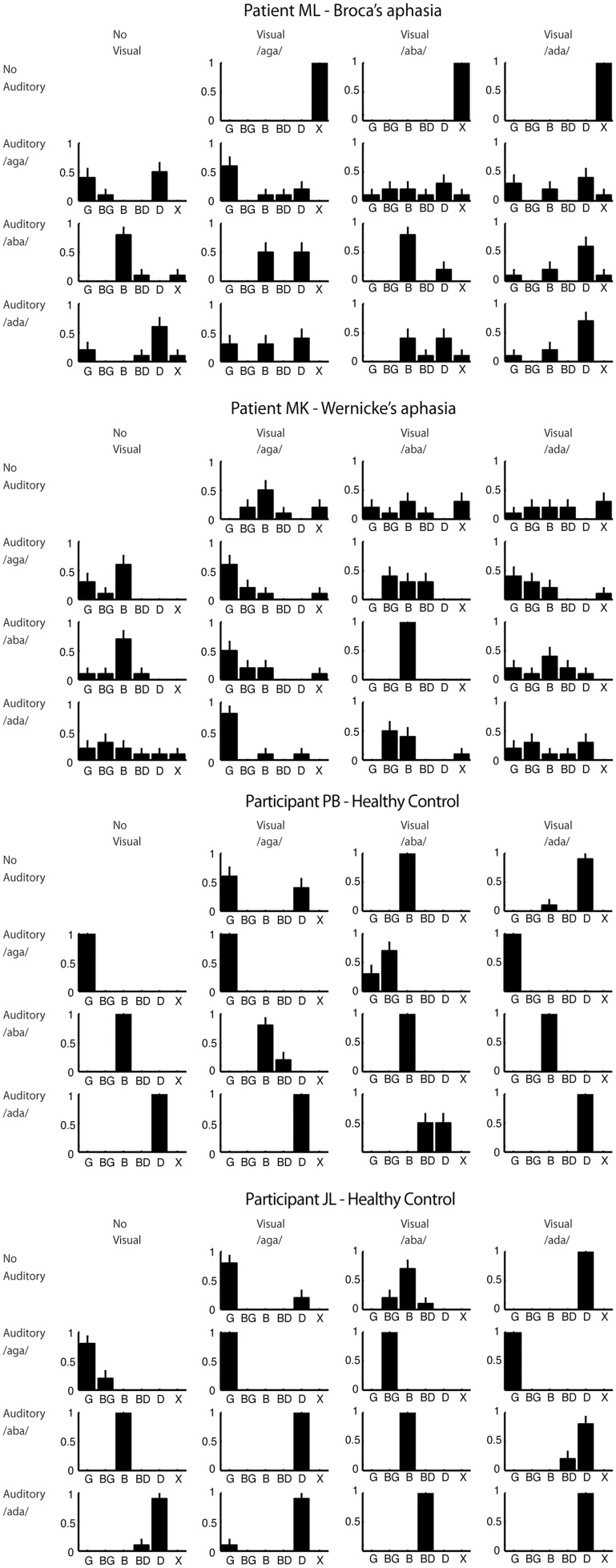
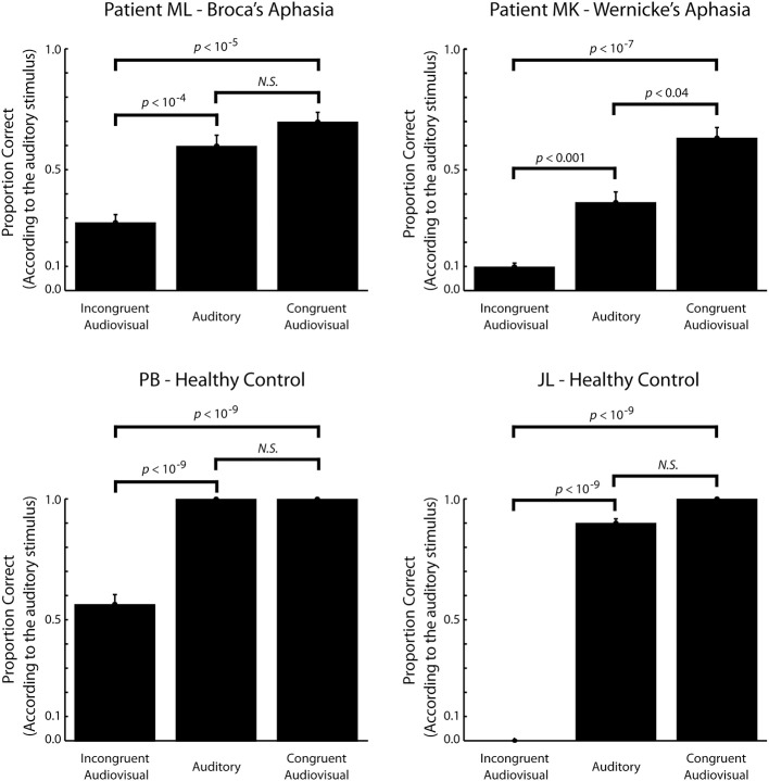
Lesions to Broca's area cause aphasia characterized by a severe impairment of the ability to speak, with comparatively intact speech perception. However, some studies have found effects on speech perception under adverse listening conditions, indicating that Broca's area is also involved in speech perception. While these studies have focused on auditory speech perception other studies have shown that Broca's area is activated by visual speech perception. Furthermore, one preliminary report found that a patient with Broca's aphasia did not experience the McGurk illusion suggesting that an intact Broca's area is necessary for audiovisual integration of speech. Here we describe a patient with Broca's aphasia who experienced the McGurk illusion. This indicates that an intact Broca's area is not necessary for audiovisual integration of speech. The McGurk illusions this patient experienced were atypical, which could be due to Broca's area having a more subtle role in audiovisual integration of speech. The McGurk illusions of a control subject with Wernicke's aphasia were, however, also atypical. This indicates that the atypical McGurk illusions were due to deficits in speech processing that are not specific to Broca's aphasia.
Keywords: Broca's area; aphasia; audiovisual; multisensory integration; speech perception.
Figures
References
-
- Andersen T. S., Tiippana K., Laarni J., Kojo I., Sams M. (2009). The role of visual spatial attention in audiovisual speech perception. Speech Commun. 51, 184–193 10.1016/j.specom.2008.07.004 - DOI
-
- Andersen T. S., Tiippana K., Lampinen J., Sams M. (2001). Modeling of audiovisual speech perception in noise, in International Conference on Auditory–Visual Speech Processing (AVSP) (Aalborg: ), 172–176.
LinkOut - more resources
Full Text Sources
Other Literature Sources