. 2015 Apr 29:6:396.
doi: 10.3389/fmicb.2015.00396. eCollection 2015.

Host cell type-dependent translocation and PhoP-mediated positive regulation of the effector SseK1 of Salmonella enterica

Affiliations

Host cell type-dependent translocation and PhoP-mediated positive regulation of the effector SseK1 of Salmonella enterica

Fernando Baisón-Olmo et al. Front Microbiol. .

Abstract

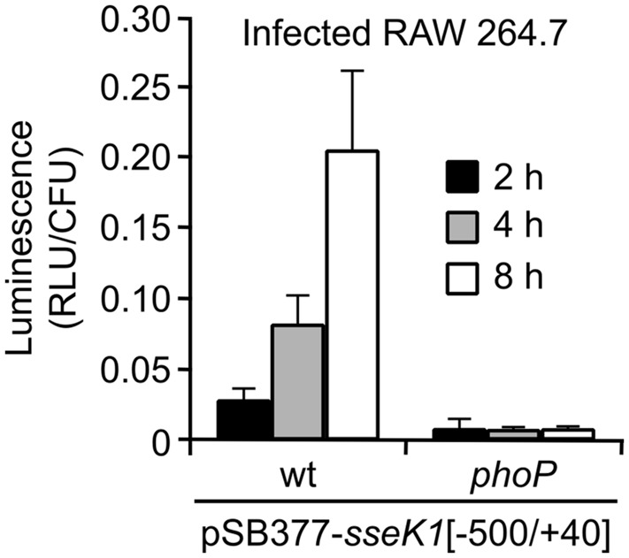
Salmonella enterica expresses two virulence-related type III secretion systems (T3SSs) encoded in Salmonella pathogenicity island 1 (SPI1) and SPI2, respectively. SseK1 is a poorly characterized substrate of the SPI2-encoded T3SS. Here, we show that this effector is essential to get full virulence both in oral and intraperitoneal mice infections, in spite of not having a role in invasion or intracellular proliferation in cultured mammalian cells. In vitro, expression of sseK1 was higher in media mimicking intracellular conditions, when SPI2 was induced, but it was also significant under SPI1 inducing conditions. A detailed analysis of translocation of SseK1 into host cells unveiled that it was a substrate of both, T3SS1 and T3SS2, although with different patterns and kinetics depending on the specific host cell type (epithelial, macrophages, or fibroblasts). The regulation of the expression of sseK1 was examined using lacZ and bioluminescent lux fusions. The two-component system PhoQ/PhoP is a positive regulator of this gene. A combination of sequence analysis, directed mutagenesis and electrophoretic mobility shift assays showed that phosphorylated PhoP binds directly to the promoter region of sseK1 and revealed a PhoP binding site located upstream of the predicted -35 hexamer of this promoter.

Keywords: PhoQ/PhoP two-component system; Salmonella; SseK1; bioluminescence; epithelial cells; fibroblasts; macrophages; type III secretion.


Figures

FIGURE 1
FIGURE 1
Competitive index (CI) analysis for an sseK1 null mutant. (A) Graphical representation of CI analysis of a strain carrying a mutation in sseK1 after intraperitoneal (ip) and oral mice infections. (B) Analysis of invasion of the sseK1 mutant in mixed infections with a trg::MudJ mutant used as the wild-type strain. (C) Analysis of intracellular proliferation of the sseK1 mutant in mixed infections with a trg::MudJ mutant used as the control strain. The CIs are the means from three infections. Error bars represent the SD. wt, wild-type strain. Asterisks denote that the CIs are significantly different from 1 for a t-test P value < 0.05.
FIGURE 2
FIGURE 2
Expression of sseK1 in different culture media. β-Galactosidase activities were measured from overnight cultures of a Salmonella enterica strain carrying a chromosomal sseK1::lacZ translational fusion. (A) Bacteria were incubated overnight at 37°C in LB with 0.3 M NaCl without shaking (SPI1) or in LPM with shaking (SPI2). (B) Different concentrations of NaCl were used to test the role of osmolarity on sseK1 expression under SPI1-inducing conditions. (C) The effects of pH, oxygen limitation and butyrate on sseK1 expression were tested in LB. (D) Activities were measured after growth in LPM with different pH and Mg++ concentrations, as indicated. Means and SD from three independent β-galactosidase measurements are shown. Statistical significance is shown by asterisks representing t-test P values: P < 0.05; ∗∗P < 0.01. C: reference for statistical comparison.
FIGURE 3
FIGURE 3
Translocation of SseK1 into mammalian cells. Human epithelial HeLa cells (A,B), RAW264.7 murine macrophage-like cells (C,D) and NRK-49F normal rat kidney fibroblasts (E,F) were infected with derivatives of S. enterica serovar Typhimurium 14028 (wild-type, wt, ΔSPI1, ΔSPI2 and ΔSPI1 ΔSPI2 strains) carrying a plasmid expressing an SseK1-CyaA’ fusion from a constitutive promoter (A,C,E) or a chromosomal SseK1-CyaA’ fusion expressed under the native sseK1 promoter (B,D,F). Bacteria were grown under SPI1-inducing conditions for most infections. Non-invasive bacteria were used specifically for infections of RAW264.7 cells for 4, 8, and 16 h. To detect translocation, the level of cAMP was measured 1, 2, 4, 8, and 16 h p.i. Means and SD from triplicate experiments are represented.
FIGURE 4
FIGURE 4
Positive regulation of the expression of sseK1 by the PhoQ/PhoP system. β-Galactosidase activities were measured from SPI1-inducing (A) and SPI2-inducing (B) cultures of several S. enterica serovar Typhimurium strains: wild-type 14028 (wt), null mutants (hilA, hilD, ssrB, rcsB, and dam), and mutants with constitutive activation of the PhoQ/PhoP system and the Rcs system, respectively (phoQ24 and rcsC54), carrying an sseK1::lacZ translational fusion. The role of SsrB in the regulation by PhoP was investigated in a double mutant phoQ24 ssrB. Means and SD from three independent β-galactosidase measurements are shown. The effect of the PhoQ/PhoP system (C) and of SsrB (D) on sseK1 expression at the protein level was assessed by immunoblot analysis using strains expressing SseK1-3xFLAG. A monoclonal anti-FLAG antibody was used to detect the fusion protein and a polyclonal anti-DnaK antibody was used to get a loading control. Representative gels are shown together with quantification of bands (SseK1/DnaK ratio) from duplicate gels. Statistical significance of the differences between wt and mutant strains is shown by asterisks representing t-test P values: P < 0.05; ∗∗P < 0.01.
FIGURE 5
FIGURE 5
Identification of a PhoP box in the promoter of sseK1. (A) Analysis of the promoter region of sseK1. The sequence surrounding the transcriptional start site (+1) is shown. The start of the coding sequence, the putative ribosomal binding site and the consensus sequences for σ70-dependent transcription (-10 and -35) are indicated. Putative PhoP-binding motifs are marked in pink and a putative PhoP-box is underlined with an arrow. A fragment of DNA containing the promoter region and 5 untranslated region of sseK1 (-500/+40) was inserted into plasmid pIC552 to generate a lacZ transcriptional fusion (B) and into plasmid pSB377 to generate a luxCDABE transcriptional fusion (C). These plasmids and the corresponding original empty vectors were introduced into S. enterica serovar Typhimurium strain 14028 (wt) or a phoP-null mutant, and β-galactosidase activities or luminescence, respectively, were measured in cultures grown to stationary phase in liquid LPM at pH 5.8. Luminescence was also measured from the wt, phoP and phoQ24 strains carrying derivatives of pSB377 with the promoter region of sseK1 or variants with the indicated mutations and grown under SPI1 (D) or SPI2 (E) inducing conditions. RLU: relative light units.
FIGURE 6
FIGURE 6
Direct interaction of phosphorylated PhoP with the promoter region of sseK1. Purified His6-PhoP was phosphorylated in vitro with acetyl phosphate. (A) DNA fragments containing the promoter regions of sseK1 (-500/+40), phoN and slyB were PCR amplified using fluorochrome-labeled primers and incubated with the indicated concentrations of phosphorylated His6-PhoP (PhoP-P). Electrophoretic mobility shift assays were used to detect binding. (B) DNA fragments containing the promoter regions of sseK1 (-300/-1) wild-type (wt) or with mutations T– > C at positions -73, -72, -62 y -61 (PhoP box mutant) were PCR amplified using fluorochrome-labeled primers and incubated with the indicated concentrations of phosphorylated His6-PhoP (PhoP-P). Electrophoretic mobility shift assays were used to detect binding.
FIGURE 7
FIGURE 7
Intracellular PhoP-dependent expression of sseK1. Two strains of S. enterica serovar Typhimurium (wild-type, wt, and phoP mutant) carrying a plasmid expressing an sseK1::luxCDABE transcriptional fusion (pSB377-sseK1[-500/+40]) were grown for 24 h in LB at 37°C with aeration (non-invasive conditions). These bacteria were used to infect RAW264.7 murine macrophage-like cells and luminescence produced by intracellular bacteria was measured 2, 4, and 8 h p.i.

Similar articles

Cited by

References

    1. Aiastui A., Pucciarelli M. G., García-Del Portillo F. (2010). Salmonella enterica serovar typhimurium invades fibroblasts by multiple routes differing from the entry into epithelial cells. Infect. Immun. 78 2700–2713 10.1128/IAI.01389-09 - DOI - PMC - PubMed
    1. Baisón-Olmo F., Cardenal-Muñoz E., Ramos-Morales F. (2012). PipB2 is a substrate of the Salmonella pathogenicity island 1-encoded type III secretion system. Biochem. Biophys. Res. Commun. 423 240–246 10.1016/j.bbrc.2012.05.095 - DOI - PubMed
    1. Bajaj V., Hwang C., Lee C. A. (1995). hilA is a novel ompR/toxR family member that activates the expression of Salmonella typhimurium invasion genes. Mol. Microbiol. 18 715–727 10.1111/j.1365-2958.1995.mmi_18040715.x - DOI - PubMed
    1. Bajaj V., Lucas R. L., Hwang C., Lee C. A. (1996). Co-ordinate regulation of Salmonella typhimurium invasion genes by environmental and regulatory factors is mediated by control of hilA expression. Mol. Microbiol. 22 703–714 10.1046/j.1365-2958.1996.d01-1718.x - DOI - PubMed
    1. Bakowski M. A., Braun V., Brumell J. H. (2008). Salmonella-containing vacuoles: directing traffic and nesting to grow. Traffic 9 2022–2031 10.1111/j.1600-0854.2008.00827.x - DOI - PubMed

LinkOut - more resources