. 2015 Feb 1;8(2):1622-30.
eCollection 2015.

ClC-3 chloride channel modulates the proliferation and migration of osteosarcoma cells via AKT/GSK3β signaling pathway

Affiliations

ClC-3 chloride channel modulates the proliferation and migration of osteosarcoma cells via AKT/GSK3β signaling pathway

Shuai Du et al. Int J Clin Exp Pathol. .

Abstract

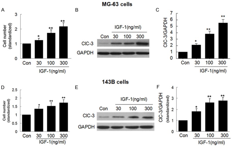
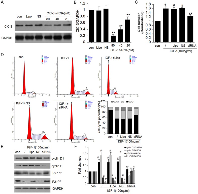
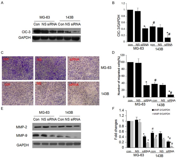
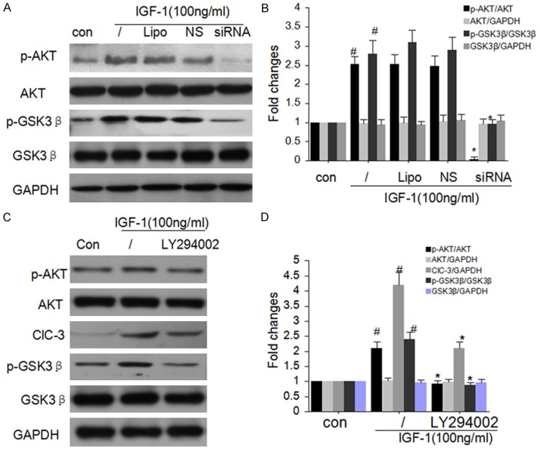
In cultured human osteosarcoma (OS) cells, we recently demonstrated that insulin-like growth factors (IGF-1)-induced MG-63 and 143B human OS cells proliferation were consistent with increasing ClC-3 expression, and ClC-3 was up-regulated in cells with high metastatic potency. Blockade of ClC-3 greatly suppressed the phosphorylation activation of Akt/GSK3β. We also found that blockade of ClC-3 effectively down-regulated the expression of cyclin D1 and cyclin E, and caused activation of p27(KIP) and p21(CIP). The synthesized effects on these proteins which play a major role in cell cycle regulation bring about G0/G1 cell cycle arrest in MG-63 cells, and finally abrogate the cell proliferation. Besides, ClC-3 deletion attenuates OS cell migration via down-regulation the expression of MMP-2 and MMP-9. Such information suggests that ClC-3 might be a potential target for anti-OS.

Keywords: AKT/GSK3β signaling pathway; ClC-3 chloride channel; osteosarcoma; proliferation and migration.


Figures

Figure 1
Figure 1
ClC-3 expression parallels with OS cells proliferation. A. Effect of 30-300 ng/ml IGF-1 on the cell viability in MG-63 cells. B and C. Effect of 30-300 ng/ml IGF-1 on the expression of ClC-3 in MG-63 cells. (Data represent mean ± SD (n = 4), Con: control, *P < 0.05 vs. con; **P < 0.01 vs. con. ANOVA test was used). D. Effect of 30-300 ng/ml IGF-1 on the cell viability in 143B cells. E and F. Effect of 30-300 ng/ml IGF-1 on the expression of ClC-3 in 143B cells. (Data represent mean ± SD (n = 4), Con: control, *P < 0.05 vs. con; **P < 0.01 vs. con. ANOVA test was used).
Figure 2
Figure 2
Effect of ClC-3 siRNA on cell cycle in MG-63 cells. A and B. Effect of 20-80 nM ClC-3 siRNA on the expression of endogenous ClC-3 protein in MG-63 cells detected by western blot analysis. (Data represent mean ± SD (n = 4), con: control, Lipo: Liposomal, NS: Negative siRNA, **P < 0.01 vs. con. ANOVA test was used). C. Knock-down of ClC-3 reduces the IGF-1-induced cell viability in MG-63 cells. D. Knock-down of ClC-3 arrests cell cycle in G0/G1 phase in MG-63 cells. E and F. ClC-3 siRNA restrained cyclin D1, cyclin E and enhanced p21, p27 protein expression induced by 100 ng/ml IGF-1 respectively in MG-63 cells. (Data represent mean ± SD (n = 4), con: control, Lipo: Liposomal, NS: Negative siRNA, #P < 0.05 vs. con; **P < 0.01 vs. IGF-1 only. ANOVA test was used).
Figure 3
Figure 3
ClC-3 knockdown attenuate cell migration in OS cells. A and B. The endogenous expression of ClC-3 in cells with different metastatic potency (MG-63 and 143B) and effect of ClC-3 siRNA on the expression of ClC-3 protein in both cells detected by western blot analysis. (Data represent mean ± SD (n = 4), con: control, NS: Negative siRNA, *P < 0.05 vs. MG-63 con; #P < 0.05 vs. MG-63 con; *#P < 0.05 vs. 143B con. ANOVA test was used). C and D. ClC-3 knockdown attenuated the migration ability of MG-63 and 143B cells detected by transwell assay. (Data represent mean ± SD (n = 4), con: control, NS: Negative siRNA, *P < 0.05 vs. MG-63 con; #P < 0.05 vs. MG-63 con; *#P < 0.05 vs. 143B con. ANOVA test was used). E and F. ClC-3 knockdown suppressed expression of MMP-2 and MMP-9 in MG-63 and 143B cells. (Data represent mean ± SD (n = 4), con: control, NS: Negative siRNA, *P < 0.05 vs. MG-63 con; #P < 0.05 vs. MG-63 con; *#P < 0.05 vs. 143B con. ANOVA test was used).
Figure 4
Figure 4
ClC-3 knockdown attenuates Akt signaling. A and B. ClC-3 knockdown significantly attenuated the phosphorylations of GSK-3β and Akt detected by western blot analysis. (Data represent mean ± SD (n = 4), con: control, Lipo: Liposomal, NS: Negative siRNA, #P < 0.05 vs. con; *P < 0.05 vs. IGF-1 only. ANOVA test was used). C and D. Akt inhibitor LY294002 diminished the expression of ClC-3 and the phosphorylations of GSK-3β. (Data represent mean ± SD (n = 4), con: control, Lipo: Liposomal, NS: Negative siRNA, #P < 0.05 vs. con; *P < 0.05 vs. IGF-1 only. ANOVA test was used).

References

    1. Adhikari AS, Agarwal N, Wood BM, Porretta C, Ruiz B, Pochampally RR, Iwakuma T. CD117 and Stro-1 identify osteosarcoma tumor-initiating cells associated with metastasis and drug resistance. Cancer Res. 2010;70:4602–4612. - PMC - PubMed
    1. Brennecke P, Arlt MJ, Campanile C, Husmann K, Gvozdenovic A, Apuzzo T, Thelen M, Born W, Fuchs B. CXCR4 antibody treatment suppresses metastatic spread to the lung of intratibial human osteosarcoma xenografts in mice. Clin Exp Metastasis. 2014;31:339–349. - PMC - PubMed
    1. Hirotsu M, Setoguchi T, Sasaki H, Matsunoshita Y, Gao H, Nagao H, Kunigou O, Komiya S. Smoothened as a new therapeutic target for human osteosarcoma. Mol Cancer. 2010;9:5. - PMC - PubMed
    1. Guan YY, Wang GL, Zhou JG. The ClC-3 Cl-channel in cell volume regulation, proliferation and apoptosis in vascular smooth muscle cells. Trends Pharmacol Sci. 2006;27:290–296. - PubMed
    1. Li M, Wu DB, Wang J. Effects of volume-activated chloride channels on the invasion and migration of human endometrial cancer cells. Eur J Gynaecol Oncol. 2013;34:60–4. - PubMed