Crescentic acute glomerulonephritis with isolated C3 deposition: a case report and review of literature
- PMID: 25973075
- PMCID: PMC4396300
Crescentic acute glomerulonephritis with isolated C3 deposition: a case report and review of literature
Abstract
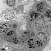
An eight-year-old girl, presenting with palpebral edema, gross hematuria, and foam in urine, was admitted to our hospital. Investigations indicated increased serum antistreptolysin O (ASO) and anti-mycoplasma antibody titers. Renal biopsy showed crescentic poststreptococcal acute glomerulonephritis (CPAGN) with isolated C3 deposition in the glomeruli. Electro-microscope examination showed subepithelial deposition of electron dense material. She received the double pulse therapies of methylprednisolone and cyclophosphamide as well as the treatment of oral prednisolone, angiotensin converting enzyme-II (ACE-II) inhibitor, dipyridamole and traditional Chinese medicine. The complete clinical remission was achieved after 9 months. No serious adverse effects were observed during the follow-up. Our findings indicated that CPAGN with isolated C3 deposition might have a favorable prognosis after aggressive immunosuppressive treatment. However, the influence of isolated C3 deposition on CPAGN prognosis remains to be clarified.
Keywords: C3; Crescentic poststreptococcal acute glomerulonephritis; deposition.
Figures
Similar articles
-
Membranoproliferative glomerulonephritis and C3 glomerulonephritis: frequency, clinical features, and outcome in children.Nephrology (Carlton). 2015 Apr;20(4):286-92. doi: 10.1111/nep.12382. Nephrology (Carlton). 2015. PMID: 25524631
-
[Crescentic and necrotizing glomerulonephritis with C3 deposition].Nihon Jinzo Gakkai Shi. 2008;50(1):51-8. Nihon Jinzo Gakkai Shi. 2008. PMID: 18318244 Japanese.
-
A case of C3 glomerulonephritis successfully treated with eculizumab.Pediatr Nephrol. 2015 Jun;30(6):1033-7. doi: 10.1007/s00467-015-3061-2. Epub 2015 Mar 22. Pediatr Nephrol. 2015. PMID: 25796589
-
Acute poststreptococcal glomerulonephritis: the most common acute glomerulonephritis.Pediatr Rev. 2015 Jan;36(1):3-12; quiz 13. doi: 10.1542/pir.36-1-3. Pediatr Rev. 2015. PMID: 25554106 Review.
-
Membranoproliferative glomerulonephritis with isolated C3 deposits: case report and literature review.Ultrastruct Pathol. 2011 Feb;35(1):42-6. doi: 10.3109/01913123.2010.532902. Ultrastruct Pathol. 2011. PMID: 21265634 Review.
Cited by
-
Mycoplasma pneumoniae Infection Associated C3 Glomerulopathy Presenting as Severe Crescentic Glomerulonephritis.Case Rep Nephrol. 2021 Sep 27;2021:6295543. doi: 10.1155/2021/6295543. eCollection 2021. Case Rep Nephrol. 2021. PMID: 34616577 Free PMC article.
-
Angiotensin II and post-streptococcal glomerulonephritis.Clin Exp Nephrol. 2024 May;28(5):359-374. doi: 10.1007/s10157-023-02446-7. Epub 2024 Jan 3. Clin Exp Nephrol. 2024. PMID: 38170299 Review.
-
Lupus nephritis or not? A simple and clinically friendly machine learning pipeline to help diagnosis of lupus nephritis.Inflamm Res. 2023 Jun;72(6):1315-1324. doi: 10.1007/s00011-023-01755-7. Epub 2023 Jun 10. Inflamm Res. 2023. PMID: 37300586 Free PMC article.
References
-
- Gale DP, Maxwell PH. C3 glomerulonephritis and CFHR5 nephropathy. Nephrol Dial Transplant. 2013;28:282–8. - PubMed
-
- Xiao X, Pickering MC, Smith RJ. C3 glomerulopathy: the genetic and clinical findings in dense deposit disease and C3 glomerulonephritis. Semin Thromb Hemost. 2014;40:465–71. - PubMed
-
- Elfenbein IB, Baluarte HJ, Cubillos-Rojas M, Gruskin AB, Coté M, Cornfeld D. Quantitative morphometry of glomerulonephritis with crescents. Diagnostic and predictive value. Lab Invest. 1975;32:56–64. - PubMed
Publication types
MeSH terms
Substances
LinkOut - more resources
Full Text Sources
Medical
Miscellaneous


