

Primary epithelioid angiosarcoma of the pleura: a case report and review of literature
- PMID: 25973118
- PMCID: PMC4396223
Primary epithelioid angiosarcoma of the pleura: a case report and review of literature
Abstract
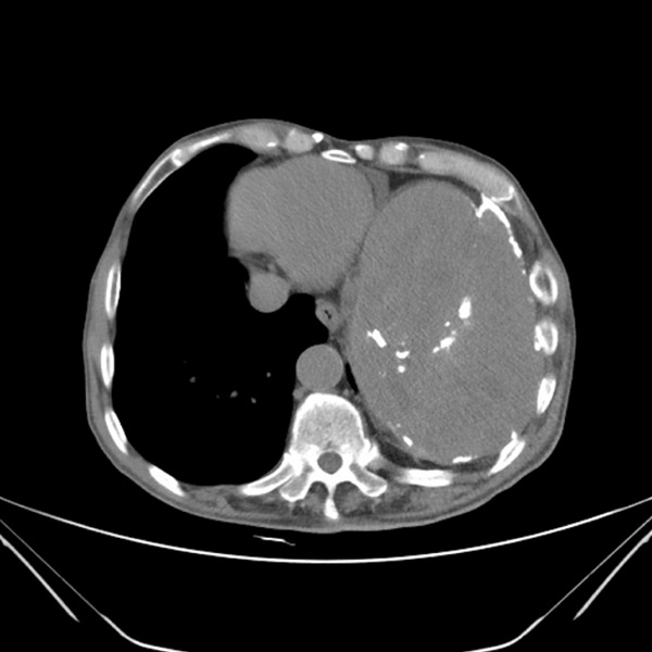
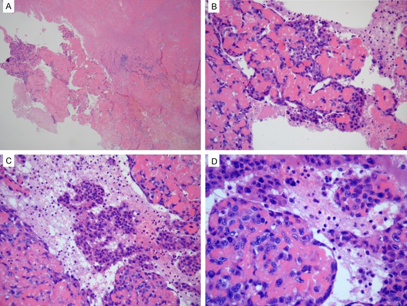
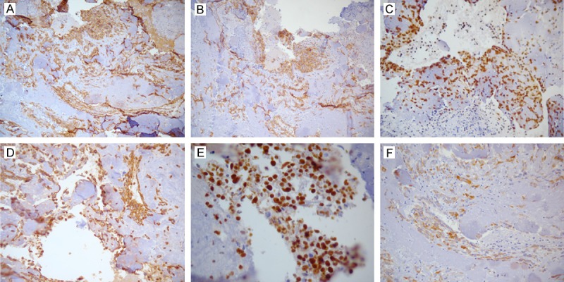
Primary angiosarcoma of the pleura is an extremely rare malignancy. Herein, we report the case of an elderly Chinese patient with primary left pleural epithelioid angiosarcoma. The 76-year-old man presented with a 4-month history of a cough with sputum expectoration and weight loss of 4 kg within one month. A chest scan showed a massive oval-shaped mass in the left pleural cavity. We then performed a left thoracotomy for tumor resection and surgical exploration. Histological examination of the resected specimen showed few viable tumor cells with significant atypia; tumor cells had large nuclei and prominent nucleoli and were arranged in a crack-like, sheeted pattern. Moreover, there was a significant amount of fibrinous exudates, hemorrhage, degeneration, and necrosis. With immunohistochemical analysis, tumor cells had strong expression of CD31, CD34, FLI-1, vimentin. Morphological and immunohistochemical findings supported the diagnosis of epithelioid angiosarcoma.
Keywords: Primary angiosarcoma; immunohistochemistry; pleura.
Figures
References
-
- Alexiou C, Clelland CA, Robinson D, Morgan WE. Primary angiosarcomas of the chest wall and pleura. Eur J Cardiothorac Surg. 1998;14:523–526. - PubMed
-
- Zhang PJ, Livolsi VA, Brooks JJ. Malignant epithelioid vascular tumors of the pleura: report of a series and literature review. Hum Pathol. 2000;31:29–34. - PubMed
-
- Kimura M, Ito H, Furuta T, Tsumoto T, Hayashi S. Pyothorax-associated angiosarcoma of the pleura with metastasis to the brain. Pathol Int. 2003;53:547–551. - PubMed
-
- Pramesh CS, Madur BP, Raina S, Desai SB, Mistry RC. Angiosarcoma of the pleura. Ann Thorac Cardiovasc Surg. 2004;10:187–190. - PubMed
Publication types
MeSH terms
LinkOut - more resources
Full Text Sources