Laparoscopic resection of intra-abdominal esophageal duplication cyst near spleen: a case report
- PMID: 25973125
- PMCID: PMC4396335
Laparoscopic resection of intra-abdominal esophageal duplication cyst near spleen: a case report
Abstract
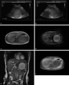
Esophageal duplication cysts (EDCs) are congenital malformations of the posterior primitive foregut and often within the thoracic esophagus. Here we describe a rare case of intra-abdominal EDC near spleen in a 20-year-old female patient with a complaint of an asymptomatic abdominal mass for 5 years. The diagnosis of intra-abdominal EDC was confirmed by the Ultrasonography (US) and Magnetic resonance imaging (MRI) as well as Histological examination. Then the patient was received the laparoscopic resection and recovered well after the operation. We conclude that the laparoscopic resection is considered to be feasible and a reasonable treatment for intra-abdominal esophageal duplication cyst.
Keywords: Laparoscopic Resection; esophageal duplication cysts (EDCs); spleen.
Figures


Similar articles
-
Laparoscopic resection of intra-abdominal esophageal duplication cyst.Surg Laparosc Endosc Percutan Tech. 2003 Jun;13(3):208-11. doi: 10.1097/00129689-200306000-00013. Surg Laparosc Endosc Percutan Tech. 2003. PMID: 12819507
-
Partial laparoscopic resection of inflamed mediastinal esophageal duplication cyst.Surg Laparosc Endosc Percutan Tech. 2007 Aug;17(4):311-2. doi: 10.1097/SLE.0b013e31805b7f26. Surg Laparosc Endosc Percutan Tech. 2007. PMID: 17710056
-
Laparoscopic excision of gastric mass yields intra-abdominal esophageal duplication cyst.Thorac Cardiovasc Surg. 2013 Sep;61(6):502-4. doi: 10.1055/s-0032-1322617. Epub 2012 Nov 21. Thorac Cardiovasc Surg. 2013. PMID: 23171952
-
Intra-abdominal esophageal duplication cysts: a review.J Gastrointest Surg. 2007 Jun;11(6):773-7. doi: 10.1007/s11605-007-0108-0. J Gastrointest Surg. 2007. PMID: 17562119 Review.
-
Anesthetic management of a patient with a mediastinal foregut duplication cyst: a case report.AANA J. 2005 Feb;73(1):55-61. AANA J. 2005. PMID: 15727285 Review.
Cited by
-
Esophageal Duplication Cysts in 97 Adult Patients: A Systematic Review.World J Surg. 2022 Jan;46(1):154-162. doi: 10.1007/s00268-021-06325-8. Epub 2021 Oct 9. World J Surg. 2022. PMID: 34628532
-
Minimally invasive surgery for adult oesophageal duplication cysts: Clinical profile and outcomes of treatment from a tertiary care centre and a review of literature.J Minim Access Surg. 2021 Oct-Dec;17(4):525-531. doi: 10.4103/jmas.JMAS_137_20. J Minim Access Surg. 2021. PMID: 34558428 Free PMC article.
-
Laparoscopic resection of an intra-abdominal esophageal duplication cyst in the ileum: a case report.Surg Case Rep. 2022 Dec 9;8(1):219. doi: 10.1186/s40792-022-01576-6. Surg Case Rep. 2022. PMID: 36484876 Free PMC article.
-
An epigastric mass? And what if it was an intra-abdominal esophageal duplication cyst: Case report.Int J Surg Case Rep. 2023 May;106:108295. doi: 10.1016/j.ijscr.2023.108295. Epub 2023 May 5. Int J Surg Case Rep. 2023. PMID: 37156202 Free PMC article.
References
-
- Herbella FA, Tedesco P, Muthusamy R, Patti MG. Thoracoscopic resection of esophageal duplication cysts. Dis Esophagus. 2006;19:132–134. - PubMed
-
- Azzie G, Beasley S. Diagnosis and Treatment of Foregut Duplications. Semin Pediatr Surg. 2003;12:46–54. - PubMed
-
- Kin K, Iwase K, Higaki J, Yoon HE, Mikata S, Miyazaki M, Imakita M, Kamiike W. Laparoscopic Resection of Intra-abdominal Esophageal Duplication Cyst. Surgical Laparoscopy Endoscopy Percutaneous Techniques. 2003;13:208–211. - PubMed
-
- Martin ND, Kim JC, Verma SK, Rubin R, Mitchell DG, Bergin D, Yeo CJ. Intra-abdominal esophageal duplication cysts: a review. J Gastrointest Surg. 2007;11:773–777. - PubMed
Publication types
MeSH terms
LinkOut - more resources
Full Text Sources
